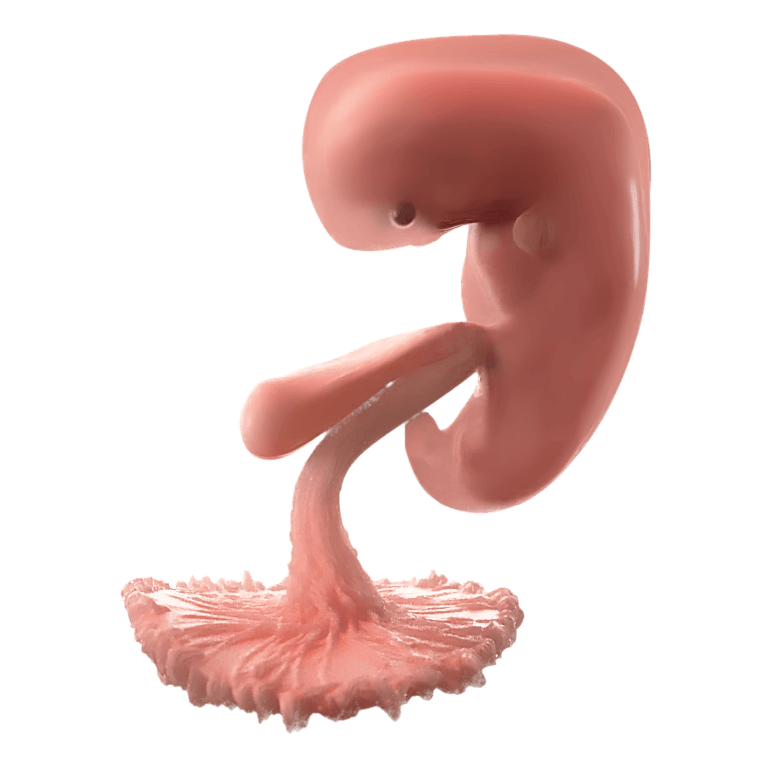
The heart is the first organ to form in a developing baby. The heart starts beating in a steady rhythm 22 days after conception, or 5 weeks and 1 day gestation.1 2 From that day on, this same heart will not stop beating until the end of that person’s life.3 By 24 days after conception, the heartbeat can be measured using a pelvic ultrasound in many viable pregnancies.4 Over the next four weeks, the average heart rate rises from an average of 110 beats per minute at the beginning of 6 weeks gestation to 174 beats per minute at the start of 9 weeks gestation, before stabilizing at a lower rate, around 150 beats per minute at 12 weeks.5 6 7 8
Become A Defender of Life
Your donation helps us continue to provide world-class research in defense of life.
DONATECharlotte Lozier Institute
Phone: 202-223-8073
Fax: 571-312-0544
2776 S. Arlington Mill Dr.
#803
Arlington, VA 22206
Highlights of the Early Heart
By four weeks gestation, a remarkable process is already underway inside the womb. Special cells in the embryo create the first and second heart fields, two regions that will shape the human heart.9
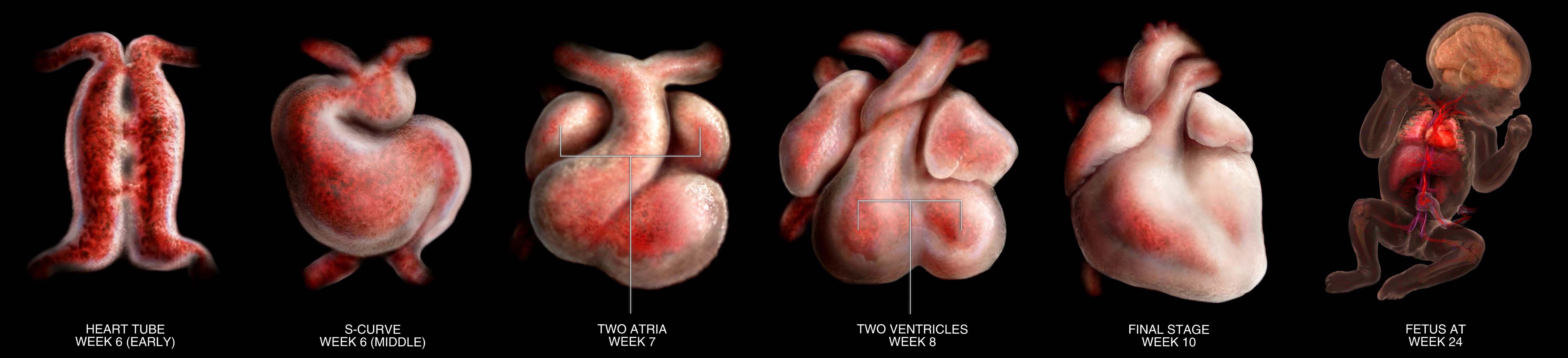
At week 5, cells from the first heart field fuse at the midline of the embryo to form a structure called the primitive heart tube. Tiny heart cells, known as primitive cardiomyocytes, begin to spontaneously twitch. These are the first flickers of a heartbeat.10 The heart tube starts to contract in a slow, wave-like motion. This gentle pulsing begins at the part of the heart where blood will enter.11 Soon these spontaneous contractions form a steady rhythm. The rhythmic heartbeat starts just 22 days after conception and will not stop until the individual dies.12
The heart tube will eventually grow into the familiar parts of the adult heart, including the left ventricle and sections of the right and left upper chambers, called atria.13 A substance called cardiac jelly, which fills the space between layers of heart tissue, provides temporary support like a soft scaffold. Cardiac jelly plays an important role in formation of early heart valves, which prevent the backflow of blood through the heart tube and assist in the forward propulsion of blood as it is pumped through the heart and out through the rest of the embryo’s body.14
Remarkably, the movement of the blood helps shape the heart. The pumping motion stretches and stimulates the growing heart cells, encouraging them to change shape and multiply in all the right places.15 During week 5, the heart begins to loop and bend, turning rightward and forming an S-shaped curve, a critical step in forming the heart’s final shape.16 During the period of cardiac looping, and the formation of heart chambers, the embryonic heart rate averages 100 to 134 beats per minute, creating shear forces that act on the developing heart cells.17
As the heart continues to grow, the tube loops and different portions expand. The heart tube dramatically increases in length thanks to newly formed heart cells that continue to join the structure. In chicks and mice the heart tube increases five-fold in length as the heart tube starts bending to the right, and different sections enlarge.18 Cells from one of the early regions called the second heart field migrate into the growing heart to help form the right ventricle, parts of the atria, the septum, which divides the heart into chambers, and the great arteries that will one day carry blood to the lungs and body.19
Between weeks 6 and 8, as the heart loops, chambers begin to emerge. Simultaneously, the walls of the ventricles form small ridges and folds that help increase strength and efficiency, in a process called trabeculation. During this time, the ventricle drastically increases in volume, as muscle cells rapidly multiply and thicken the outer heart wall. One study in mice found that ventricles increase in size nearly 100-fold!20
Meanwhile, the atria begin to form at the top of the heart. The veins feeding blood into the heart are gradually absorbed into the right atria and lined with muscle. This region, called the sinus venosus, becomes the heart’s natural pacemaker—controlling the rhythm of the early heartbeat.21
Around seven weeks, the left atrium and pulmonary veins start to form. By 8 weeks, four pulmonary veins will be connected to the left atrium.22 The pulmonary vein will carry oxygenated blood from the lungs to the heart after birth. It starts near the center of the heart but gradually shifts to become part of the left atrium thanks to the precise placement of tissue walls that grow around it.23 This careful orchestration is essential to establishing normal circulation.

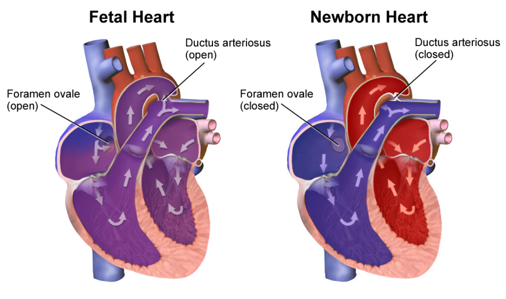
By gestational week 9, the baby’s heart is nearly complete in form, with two atria, two ventricles, and a network of vessels. However, the walls of each chamber are still growing. By 10 weeks, the baby’s heart has its characteristic four-chambered shape.24 Fetal circulation is unique; the fetal lungs and liver are bypassed since oxygen comes from the mother through the umbilical cord during pregnancy. Yet, the heart is fully functional. It beats, pumps, and supports the growing needs of the unborn child.25
Valves are needed for the baby’s heart to function properly. In the same way that car valves control an engine’s fuel, heart valves ensure blood flows in the right direction. The four-chambered heart has four valves—mitral, tricuspid, aortic, and pulmonary.
The first evidence of valve formation starts around week 6 and 7 gestation with the formation of small swellings called endocardial cushions in the early heart tube. Even at this early stage, endocardial cushions physically prevent the blood from flowing backward and guide the blood forward as it is pumped through the heart and through the rest of the embryo’s body.26
Endocardial cushions form from cardiac jelly, a gel-like material that supports the growth and pumping of the early heart.27 Specialized cells within the endocardial cushions, called endothelial cells, invade the cardiac jelly and as they move, they transform into a different cell type. This intricate cellular process called endothelial-to-mesenchymal transition is critical to formation of the heart valves.28 29 From these early structures emerge the mitral and tricuspid valves, which separate the heart’s chambers, and the aortic and pulmonary valves, which control blood leaving the heart.30 31
Once fully formed, heart valves contain tiny but strong flaps that open and close rapidly with each heartbeat. Over a lifetime, heart valves will open and close about 3 billion times faithfully directing blood flow!32
Every step of heart development demonstrates biological precision and the inherent value of life in the womb. From the moment the heart begins beating just three weeks after conception, the human heart is a working organ — pumping blood, beating rhythmically, and circulating essential nutrients through a living human being.