Search Icon
close-panel

Charlotte Lozier Institute

Phone: 202-223-8073
Fax: 571-312-0544

2776 S. Arlington Mill Dr.
#803
Arlington, VA 22206

Get Notifications

Sign up to receive email updates from Charlotte Lozier Institute.

This field is for validation purposes and should be left unchanged.

close-panel

Become A Defender of Life

Your donation helps us continue to provide world-class research in defense of life.

DONATE

Charlotte Lozier Institute

Phone: 202-223-8073
Fax: 571-312-0544

2776 S. Arlington Mill Dr.
#803
Arlington, VA 22206

Eye Formation

Dive Deeper

Few structures in the human body inspire as much wonder as the eye—a living window, built from cells that must become perfectly clear in order to see. “Perhaps biology faces no greater feat,” write one group of ophthalmologists, than building such complexity from a single cell. 1

How does the eye form?

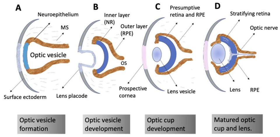
The eyes begin forming 22 days after conception, also known as 5 weeks and 1 day gestation.2 Tissue from the developing brain bulges towards the face and then folds in on itself to form an optic cup. The optic cup becomes the back of the eye, called the retina, which turns light energy into neural signals, and retinal pigment epithelium, which nourishes the retina and keeps it healthy. The optic cup also helps create the muscles that change the size of the pupil. The rim of the optic cup becomes the colorful iris, and the hole in the iris becomes the pupil.3 In week 7 retinal pigments start forming, making the eyes darker than facial skin by week 8.4

A labeled diagram illustrates four stages of Eye Formation: A) optic vesicle formation, B) optic vesicle development, C) optic cup development, and D) matured optic cup and lens, with key structures labeled in each stage.
Baby's eyes form from the interaction of nervous tissue, outer tissue called ectoderm starting in the sixth week. At birth, the newborn is born able to see, but with such low visual acuity that she is legally blind. Visual experience outside the womb helps the visual system finish developing. (Image Credit: Gregoryan)

The neural tissue connecting the developing brain to the optic cup becomes the optic nerve. As retinal ganglion cells develop, they send their axons towards the brain through the optic nerve. More and more axons grow into the nerve, filling it and widening it.5 In fact, during the second trimester, each optic nerve has over 2.8 million axons! Then the fetus and infant start reducing the number of axons, such that the adult will have 1 million axons per optic nerve. The axons that remain get coated with a fatty sheath of myelin; therefore the adult optic nerve is wider than the fetal optic nerve even though it has less axons.6

The front of the eye forms from the surface tissue. Chemical messages from the optic cup cause the surface tissue to thicken and fold in on itself creating the lens. Surrounding cells form the cornea, blood vessels, and the white part of the eyes known as the sclera.7 Chemical messages also perfectly organize collagen fibers in the lens and cornea to make them transparent. Cells in the cornea and lens actively pump out water, maintaining precise hydration and clarity critical for proper light transmission.8

Interestingly, the eyeball grows in spurts between 16 and 20 weeks and 28 and 32 weeks.9 Many have noticed that babies have large eyes compared to their faces. At birth, the eyeball is ¾ of its adult diameter and 2/3 of its adult weight.10

Perhaps biology faces no greater feat than the generation of a complex, multicellular organism from a single cell origin. The eye quintessentially represents this process through the formation of highly specific cell types, regulated by an intricate network of cellular interactions and genetic interplay of the early embryo.11
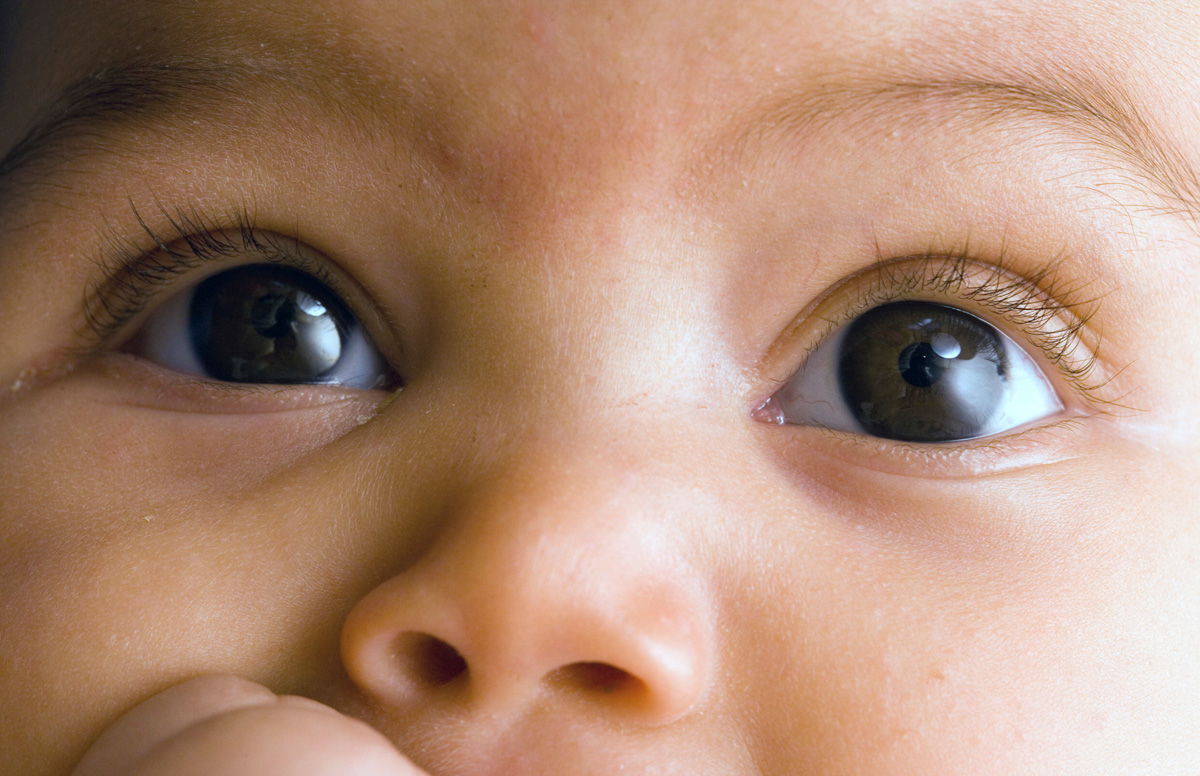
By about 23 weeks, a baby’s eye movements can reveal whether they are awake, asleep, or even potentially dreaming. (Image Credit: Science Source)(Image Credit: Science Source)
When does the fetus start moving her eyes?

Muscles start moving the eyes by week 12.12 13 Spontaneous eye movements can be detected by ultrasound starting in week 14,14 and can reveal whether a baby is awake, asleep, or dreaming starting in week 23.15 16 Between weeks 17 and 23, the fetus produces lots of different eye movements including single prolonged eye movements, and fast movements followed by slower repositioning.17 These eye movements are critical for linking the visual and motor areas of the brain before birth.18

How do photoreceptors form?

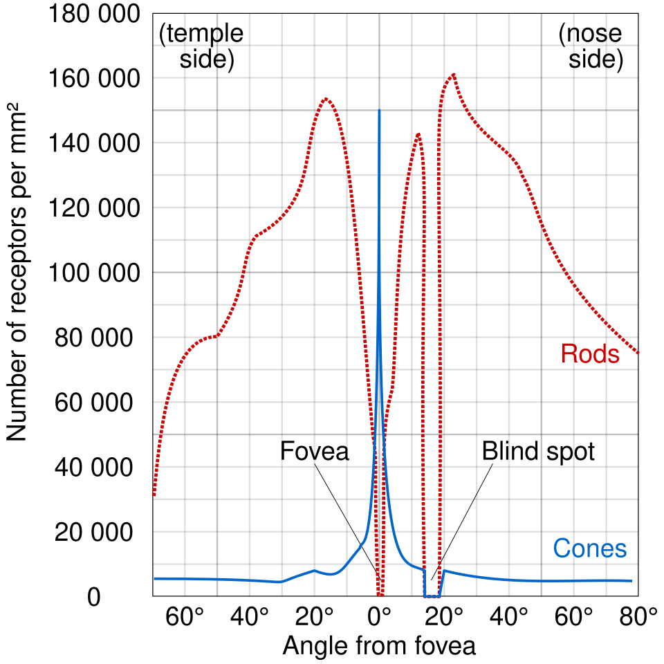
The retina, located at the back of the eye, contains two types of light-sensitive cells called rods and cones. These cells begin forming around 11 weeks.19 Rods are extremely sensitive – capable of detecting a single photon—and are responsible for seeing in dim light, while cones function best in bright light and enable color vision. In adults, each eye typically contains about 100 million rods and 6 million cones.20 While both rods and cones are formed all over the retina, cones cluster in the center, called the fovea, and rods are concentrated in the surrounding area, called the periphery.21

The organization of the retina continues to change throughout pregnancy and even after birth. Cell division in the fovea stops around 16 weeks of gestation, while cell division in the outer parts of the retina continues until about 32 weeks.22 As development proceeds, cones gradually migrate toward the center of the retina, while rods move outward—a process that continues for many months postnatally.23 Between 18 and 30 weeks, the retina overproduces relay neurons known as ganglion cells, resulting in a temporary surplus compared to the number present at birth.24

Despite their early appearance, rods are not yet fully functional; in premature infants born around 36–38 weeks, the retinal response to light is only about 2% as strong as that of an adult retina.25 Similarly, the cones continue to develop well after birth, reaching maturity around four years of age.26

The distribution of rods and cones from the outer edge (temple side) to the nose side. The center of the retina is called the fovea, and cones concentrate there. The rest of the retina is mostly covered with rods, which detect light in dim settings. The blind spot is the location on the retina where the optic nerve starts. (Image Credit: <a href="https://commons.wikimedia.org/wiki/File:Human_photoreceptor_distribution.svg">Wikimedia commons</a>)
The distribution of rods and cones from the outer edge (temple side) to the nose side. The center of the retina is called the fovea, and cones concentrate there. The rest of the retina is mostly covered with rods, which detect light in dim settings. The blind spot is the location on the retina where the optic nerve starts. (Image Credit: Wikimedia commons)
When does the fetus open her eyes?

In week eight, surface tissue interacts with the eye to form the embryo’s eyelids. Interestingly, the embryo’s eyelids actually fuse together around 10 weeks and only start separating again around 22 weeks.27 Just as some babies walk earlier than others, some premature infants open their eyes before others. Between 23 and 26 weeks the eyelids have fully separated in most fetuses and the premature baby blinks.28 29

What can the fetus see?

By about 6 months, the visual system is already responding to the world of light. Premature infants as young as 24 weeks display brain responses to flashes of light,30 31 and fetuses at 26 weeks move their eyes towards face-like patterns of light projected into the womb.32 After birth, the pupil begins to constrict and dilate to regulate the amount of light entering the eye. This pupillary reflex has first been detected in preterm babies at 30 weeks, and is consistently seen by 35 weeks.33

What can premature babies see?

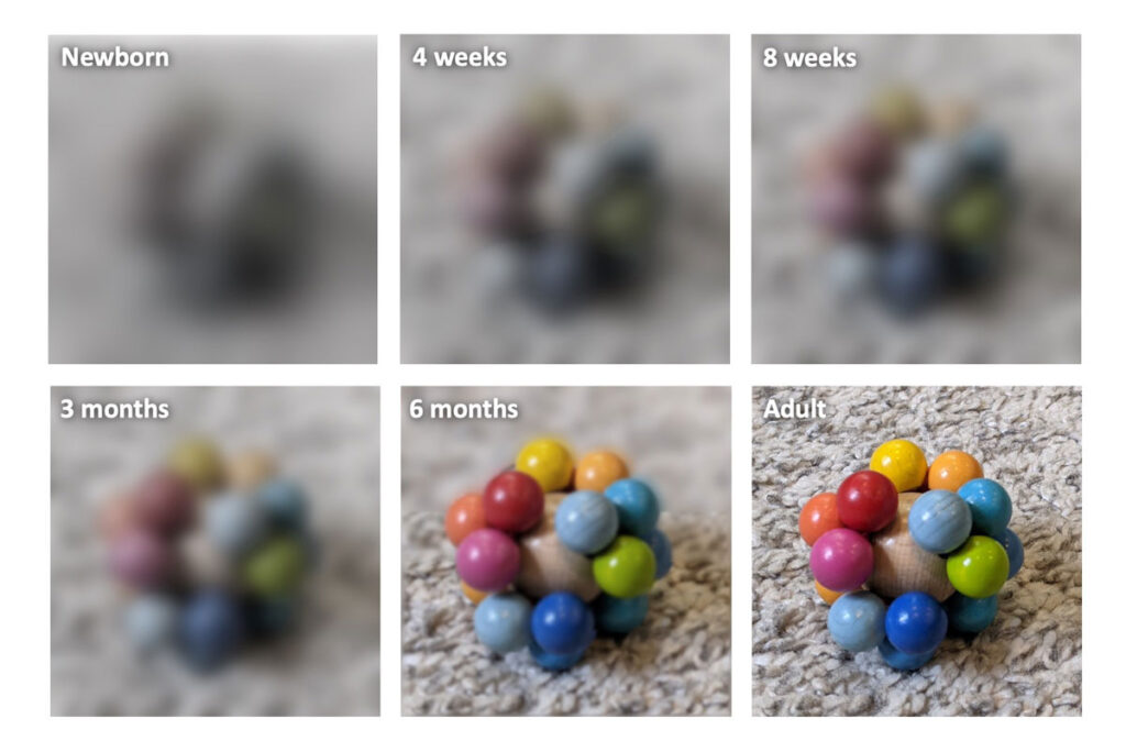
Premature infants can look directly at objects or lights starting between 30 and 32 weeks. These infants start moving their eyes to track moving objects at 34 weeks.34 Interestingly, they look preferentially at a pattern over a gray circle starting at 34 weeks. This is especially impressive because their visual acuity is around 20/2000.35 Simply put, that means that an object that an average adult could see 2000 feet away would need to be just 20 feet away for the baby to be able to see it. For perspective, a person is legally blind if their visual acuity is 20/200 or worse. Most babies born around their due date have a visual acuity of 20/866.36 Babies have adult levels of visual acuity around 3 or 4 years old.37

Major milestones in eye development:

Gestational Age Milestone
Week 6 Lens placode and optic cup form.38
Week 8 Pigments developing in retina.39 Eyelids form.40
Week 10 The eyes have all of their major components including the lens, cornea, and retina.41 Eyelids fuse together.42
Week 11 Rods and cones start forming.43
Week 12 First eye movements recorded.44
Week 14 Several eyelashes have formed.45 Spontaneous eye movements begin.46
Week 22 Eyelids start reopening.47
Week 23 Rapid eye movements begin.48
Week 24 Premature infants show brain responses to single or rhythmic flashes of light.49
Week 26 The fetus moves her eyes to look at face-like patterns projected into the womb.50 Blinking consistently observed.51
Week 28 First brain responses to light measured in utero.52
Week 30 The pupils constrict and dilate in response to light.53 Fetal eye movements are linked to brain activity in visual areas.54
Week 32 Premature infants can fixate on lights.55
Week 34 Premature infants can visually track a moving object.56
Sperm-egg fusion
central part of the retina