Search Icon
close-panel

Charlotte Lozier Institute

Phone: 202-223-8073
Fax: 571-312-0544

2776 S. Arlington Mill Dr.
#803
Arlington, VA 22206

Get Notifications

Sign up to receive email updates from Charlotte Lozier Institute.

This field is for validation purposes and should be left unchanged.

close-panel

Become A Defender of Life

Your donation helps us continue to provide world-class research in defense of life.

DONATE

Charlotte Lozier Institute

Phone: 202-223-8073
Fax: 571-312-0544

2776 S. Arlington Mill Dr.
#803
Arlington, VA 22206

Fetal vs. Neonatal Circulation

Dive Deeper
The Circulatory System Before and After Birth

Before a baby takes her first breath, her circulatory system works differently during pregnancy than it will after birth. Inside the womb, the developing baby doesn’t use her lungs or digestive system yet. Instead, oxygen and nutrients come directly from the mother through the placenta, and the baby’s heart and blood vessels are specially designed to make the most of that connection.1


The Fetal Circulatory System: Designed for the Womb

The fetal heart and circulatory system are beautifully structured to meet the needs of the baby growing in the womb. The placenta supplies oxygen and nutrients during pregnancy. Three temporary “shunts,” or shortcuts, reroute blood to ensure that oxygen-rich blood from the placenta reaches vital organs like the heart and brain. These shunts are:

  1. The ductus venosus, which lets blood bypass the liver.
  2. The foramen ovale, which lets blood flow between the right and left atria of the heart.
  3. The ductus arteriosus, which connects the pulmonary artery, heading to the lungs, directly to the aorta, carrying blood to the body.2
The fetal circulatory system includes three shunts to divert oxygenated blood from the placenta to the heart, reduce circulation in the lungs, and divert blood back to the placenta. (Image Credit: <a href="https://commons.wikimedia.org/wiki/File:2916_Fetal_Circulatory_System-02.jpg" target="_blank">OpenStax College</a>, 19 June 2023, Public Domain)
The fetal circulatory system includes three shunts to divert oxygenated blood from the placenta to the heart, reduce circulation in the lungs, and divert blood back to the placenta. (Image Credit: OpenStax College, 19 June 2023, Public Domain)

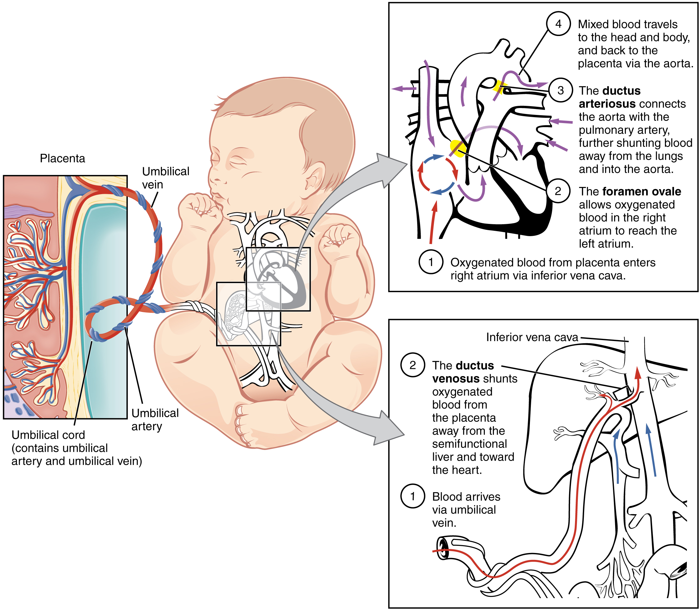
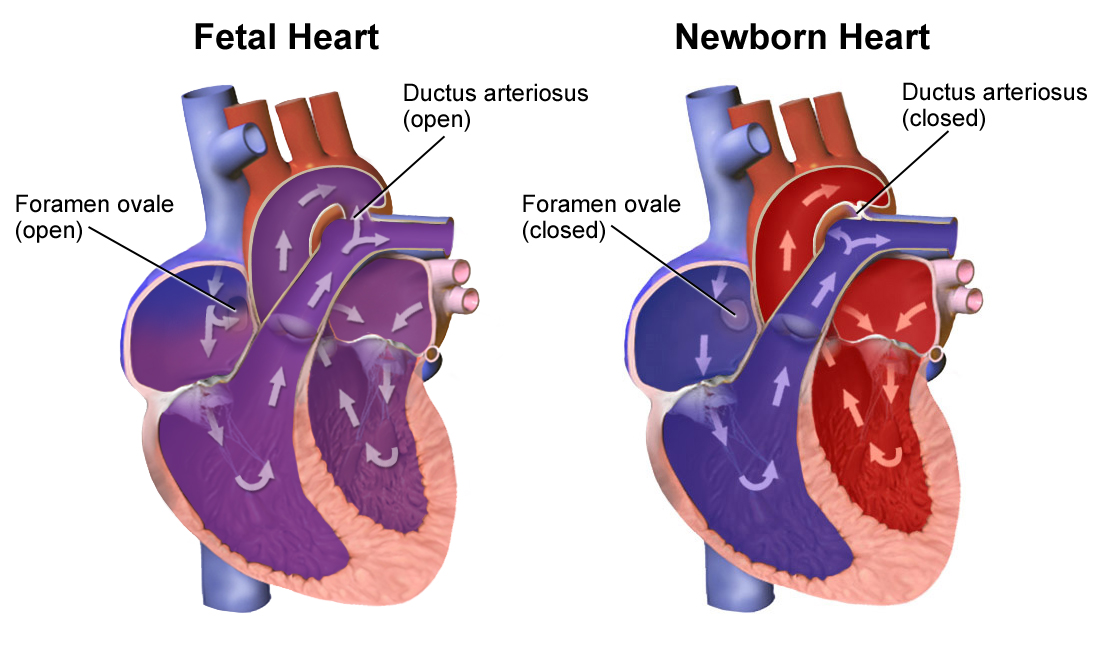
Blood from the placenta travels through the umbilical vein. About half of it skips the liver via the ductus venosus, entering a large vein that leads straight to the heart. The rest goes through the liver to nourish it. Once in the heart, the blood flows into the right atrium, but because the lungs aren’t working yet, most of the blood bypasses them through the foramen ovale, a flap-like opening into the left atrium.3

Another clever shortcut is the ductus arteriosus, which sends most of the blood that would normally go to the lungs straight into the aorta instead. This ensures oxygenated blood reaches the body without needing to pass through the fetal lungs.4

Only a small amount of blood— just 8-10%— travels through the lungs around 20 weeks gestation because the resistance in fetal lung vessels is extremely high. The returning blood, now carrying less oxygen, exits through two umbilical arteries back to the placenta, where it will be refreshed with oxygen and nutrients once again.5

A Heart Built for Two: Mom and Baby Working Together

The fetal heart beats quickly, between 110 and 170 beats per minute,6 and sends more than 40% of its output to the placenta. The left side of the heart delivers oxygenated blood to the brain and heart muscle, ensuring major organs receive what they need to develop properly. At birth, the newborn heart beat decreases to about 100-120 beats per minute.7

As pregnancy continues, the baby’s lungs prepare for life outside the womb. By 30 weeks, the amount of blood going to the lungs gradually increases to about 25%. Developing fetal tissues need lots of nutrients, and blood flows through the fetal body at more than twice the rate seen in newborns.8

At birth, the ductus arteriosus and foramen ovale close so that oxygen-poor blood is pumped to the lungs, and oxygen-rich blood is pumped to the rest of the body without the blood mixing. (Image Credit: <a href="https://en.wikipedia.org/wiki/Fetal_circulation#/media/File:Neonatal_Heart_Circulation.png" target="_blank">Blausen Medical</a>, 12 November 2015, Public Domain)
At birth, the ductus arteriosus and foramen ovale close so that oxygen-poor blood is pumped to the lungs, and oxygen-rich blood is pumped to the rest of the body without the blood mixing. (Image Credit: Blausen Medical, 12 November 2015, Public Domain)
How does blood flow change after birth?

When a baby is born, everything changes within minutes. The very first breath triggers a cascade of biological signals. Oxygen enters the lungs, causing the release of nitric oxide, a powerful natural chemical that opens blood vessels in the lungs. This causes lung pressure to drop, and blood now begins to flow freely through the baby’s lungs—just like in an adult.9

At the same time, cutting the umbilical cord removes the low-resistance flow from the placenta. The baby’s body responds with a rush of hormones, boosting the heart’s pumping power to meet the demands of breathing, feeding, and staying warm.10

The four fetal shunts now begin to shut down:

  • The foramen ovale closes as pressure builds on the left side of the heart, preventing blood from flowing backward.11 Interestingly, in about one quarter of people, the septa closing the foramen ovale never fuse, allowing for a little mixing of blood between the atria. This is called a patent foramen ovale, and most people do not know they have it because it is usually harmless.12
  • The ductus arteriosus begins to narrow and close within 1 to 4 days, a process triggered by the rise in oxygen.13
  • The ductus venosus, which went from the placenta to the heart, also stops functioning shortly after birth and disappears within about 2 weeks.14
  • The connections to and from the placenta were removed by cutting the umbilical cord.15

As a result, more blood begins flowing through the lungs and liver, and the blood now flows in the pattern we recognize in adults.16

A New Circulation Pattern Emerges

As the ductus arteriousus and foramen ovale close, the output from the ventricles equalizes. This shift is so dramatic that total cardiac output nearly doubles on the first day of life, before returning to lower levels. Furthermore, the neonatal blood pressure is much lower than that of an adult as the new circulation pattern emerges. This low blood pressure means that the young heart depends more on changes in heart rate to increase blood flow.17

By about 3 weeks after birth, lung pressure has dropped below body pressure, and the newborn heart fully transitions into its postnatal form. The left ventricle, which pumps to the body, becomes stronger and thicker as it takes over the heavy lifting of circulation, while the right ventricle adapts to its new role pumping blood through the lungs.18

From Cell Growth to Size Growth: Building a Bigger Heart

After birth, the heart grows in a new way. Before birth, heart cells multiply. But soon after delivery, they stop dividing and instead grow by increasing in size. By just one month old, a baby has almost all the heart cells they will ever have.19

From there, the heart grows by increasing in volume. Heart muscle cells grow more than 30 times larger,20 and the heart itself increases almost tenfold by early adulthood.21

Interestingly, adult heart muscle cells make up 90% of the heart’s volume but only half of its total cell count.22 While in many body tissues, cells continue to divide and replace themselves, this is not the case for the heart. Only about half of heart muscle cells will divide to replace injured cells during an adult lifespan.23 Most changes in heart size result from the growth in volume of existing cells.