close-panel

Charlotte Lozier Institute

Phone: 202-223-8073
Fax: 571-312-0544

2776 S. Arlington Mill Dr.
#803
Arlington, VA 22206

Get Notifications

Sign up to receive email updates from Charlotte Lozier Institute.

Brain Connections for Movement and Sensations

Dive Deeper
How does the brain develop?

Speaking, planning, moving, and dreaming: the activities that make us who we are originate in the brain. Billions of neurons send projections all over the brain, making trillions of neural connections.1 But how exactly does the brain develop and when does it start functioning?

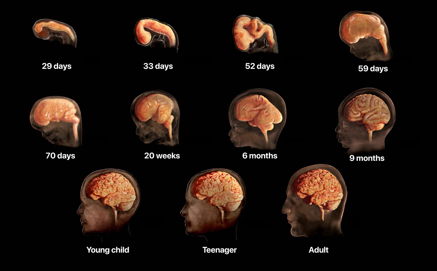
The brain and spinal cord start forming in the fifth gestational week. As the pregnancy progresses, the brain grows and folds to create distinct brain regions.
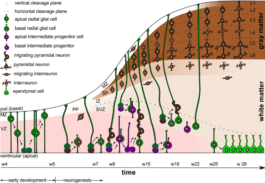
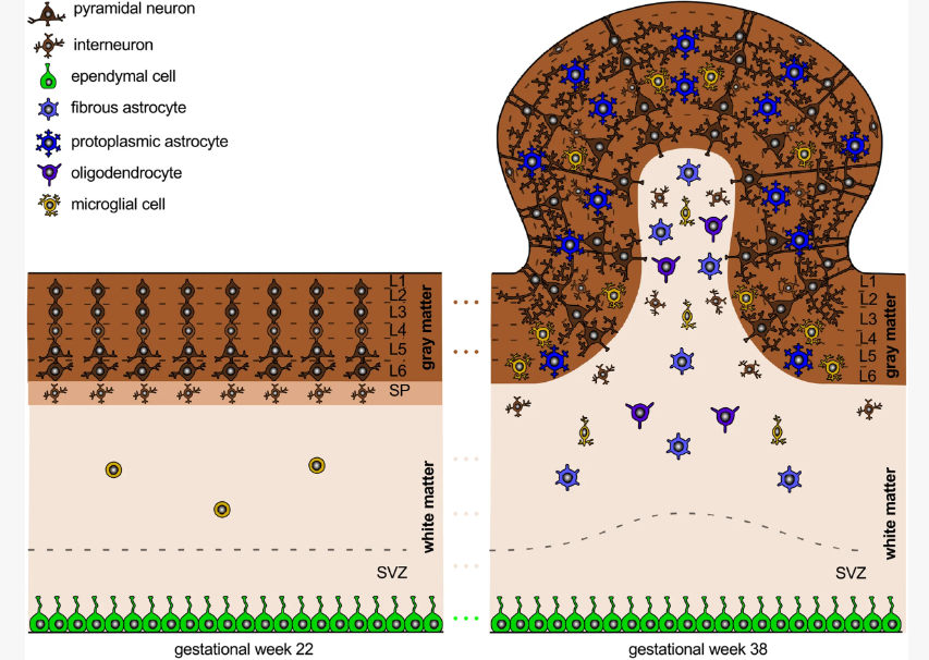
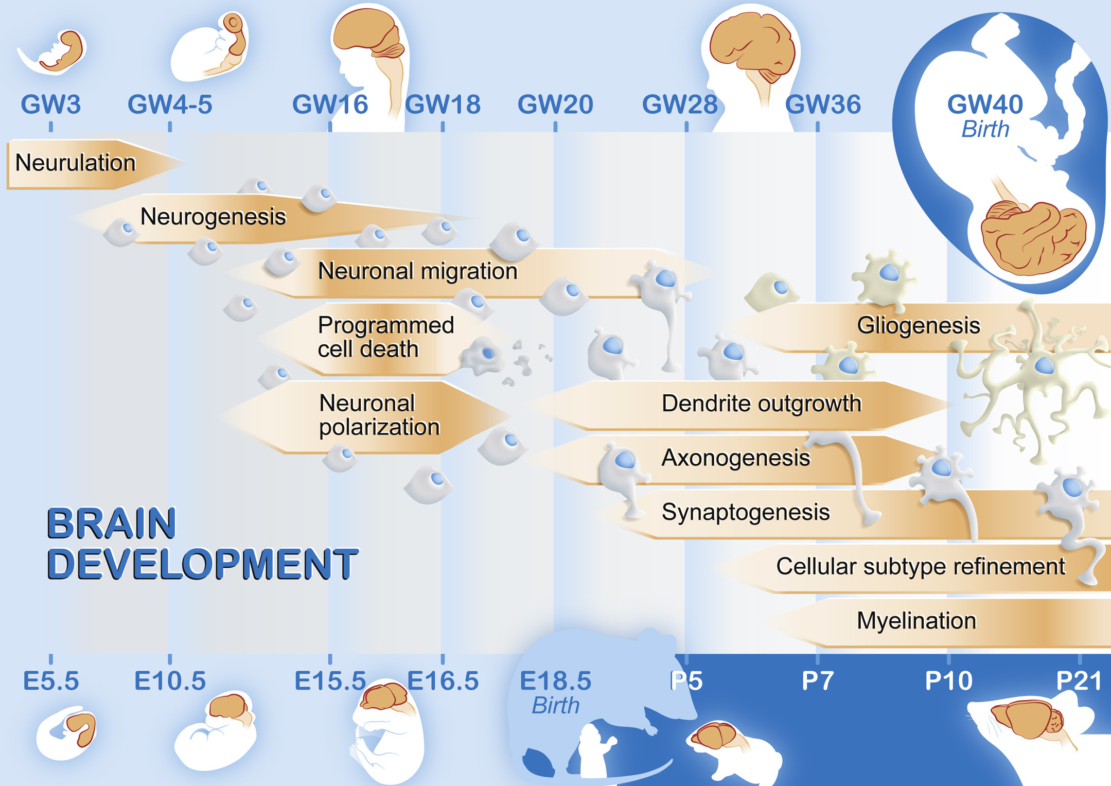
The brain assumes its adult shape around 5 ½ months.

The brain assumes its adult shape around 5 ½ months gestation. Then its smooth surface bends and folds, increasing its surface area fivefold by adding bumps and grooves.2 This allows the brain to pack in more cells. At the cellular level, neural stem cells become neurons and glia, the two basic cells of the nervous system. Most of the neurons are born near fluid-filled cavities at the center of the brain, called ventricles. Then these neurons migrate away from the center to the outer portion, called the cortex. The time of a neuron’s birth determines its position and role in the brain.3

Neurons start producing chemical messengers that allow them to communicate right away,4 and spontaneous electrical activity has been recorded from connected neurons as early as 8 weeks and 3 days gestation.5 Furthermore, reflexes require neural circuits, and the first fetal reflexes are observed before overall brain activity at 7 ½ weeks.6

7½
Weeks
Reflexes require neural circuits, and the first fetal reflexes are observed before overall brain activity at 7 ½ weeks.7
This medical visualization was created from human scanned data. The nervous system develops from the neural tube, which begins to form around the 22nd day after conception. Once the neural tube closes around the 25th day after conception, specific regions and structures in the brain begin to form and fall into place. The basics of the neural system have been established by the end of the embryonic period. Neural circuits, or connections between neurons that communicate information throughout the brain, form at a rapid rate in the fetal period. More and more brain areas become fully functional as the fetus matures and is born. The age where the brain reaches adult weight varies within the teenage years, and the brain remains a work in progress throughout adulthood. (Image Credit: Science Source)

Throughout development, the brain follows a process called blooming and pruning. Basically, the brain grows too many neurons and synaptic connections, then eliminates the connections and neurons that don’t get used frequently. This makes brain circuits more efficient. The total number of neurons peaks around 28 weeks, and the total number of synapses peaks around 2 years after birth.8

The brain will continue to undergo complex and life-long changes. The most important stages of brain maturation occur during fetal development, early childhood, and adolescence Even though brain activity has been recorded in the preborn as early as the ninth week, the frontal cortex will continue to mature well into adulthood and is not fully developed until around 25 years of age, well after birth.9

Why are the thalamus and cortex important?

Prenatal sensory experience enhances the survival of neurons involved in sensory brain circuits. In adults, sensory information enters from the eyes, ears, skin, or mouth, then goes to a relay center in the brain called the thalamus and ends in the cortex at layer 4, the layer designed to receive sensory information. Before the cortex fully matures, it grows from an area called the subplate. Neurons destined for the cortex first move into the subplate, where they wait until the cortex is sufficiently mature. In humans, the subplate forms between 12 and 17 weeks, and synapses from other brain regions enter this structure right away.10 Then the subplate neurons migrate into their mature cortical positions while keeping their synaptic connections. The subplate disappears by 6 months after birth.11

In animals, sensory information from the thalamus connects to neurons in the subplate before the sensation-receiving layer of the cortex has developed. Importantly, these connections into the subplate are organized just the way the mature cortex is organized, and function similarly as well. For instance, in brain regions that process sound, pitches of similar frequency are located adjacently in the cortex. When sounds are played to unborn animals, neurons in the subplate show frequency-organized activation as well.12 Some of the same neurons that processed sensory information in the subplate are preserved into adulthood.13 Since all of the sensory systems follow a similar pathway of development, this suggests that thalamic connections to neurons that process touch and pain in the subplate would also be active when the subplate forms.14

When does the brain start responding to sensations?

The earliest evidence of a fetus responding to sound is at 16 weeks,15 although the fine-tuning of the neurosensory pathway occurs after 20 weeks. In contrast, the fetal response to light develops late. The best direct evidence of a fetal response to a flash of light was recorded using magnetoencephalography (MEG) from fetuses recorded every two weeks from 28 weeks gestation until 2 weeks after birth.16 They found that visual responses could be detected in the fetus at 28 weeks, and that the brain response increased as the fetus got older.17 This may be because less than 10% of the light outside the womb can penetrate inside, and this light is further filtered by the fetal eyelid. Therefore, only a small amount of light reaches the fetal retina, which is mostly developed by 10 weeks.18

Sensory receptors are often in place before the entire brain circuit that governs the reaction to the sensation. For each sensory system, the first detection of receptors and evidence of their functional response is outlined in the table below:

Sensation Sensory Receptor Milestone Functional Milestone
Touch Receptors can be seen around mouth and hands at 7 ½ weeks.19 The embryo reflexively moves away at 8 weeks.20
Taste Taste buds start developing at 8 weeks and mature around 14 weeks.21 The fetus swallows different amounts of amniotic fluid based on the taste by 15 to 16 weeks.22
Hearing The cochlea starts developing at 7 weeks, but does not mature until 24 weeks.23 The fetus moves after hearing a sound by 16 weeks.24
Vision The retina has formed by 10 weeks, although individual photoreceptors like rods are not seen until 17 weeks.25 Brain responses to flashes of light were measured at 28 weeks.26
Smell Nerves from the nose to the olfactory bulb first appear at 8 weeks27 and are easily distinguishable by 10 weeks.28 The fetus distinguishes smells in the amniotic fluid by 28 weeks.29
Pain Pain receptors develop from 10 to 17 weeks.30 Fetal stress hormones increase after a painful stimulus at 18 weeks.31
Sperm-egg fusion
Sperm-egg fusion