Search Icon
close-panel

Charlotte Lozier Institute

Phone: 202-223-8073
Fax: 571-312-0544

2776 S. Arlington Mill Dr.
#803
Arlington, VA 22206

Get Notifications

Sign up to receive email updates from Charlotte Lozier Institute.

This field is for validation purposes and should be left unchanged.

close-panel

Become A Defender of Life

Your donation helps us continue to provide world-class research in defense of life.

DONATE

Charlotte Lozier Institute

Phone: 202-223-8073
Fax: 571-312-0544

2776 S. Arlington Mill Dr.
#803
Arlington, VA 22206

Ear Formation

Dive Deeper
How does the ear form?

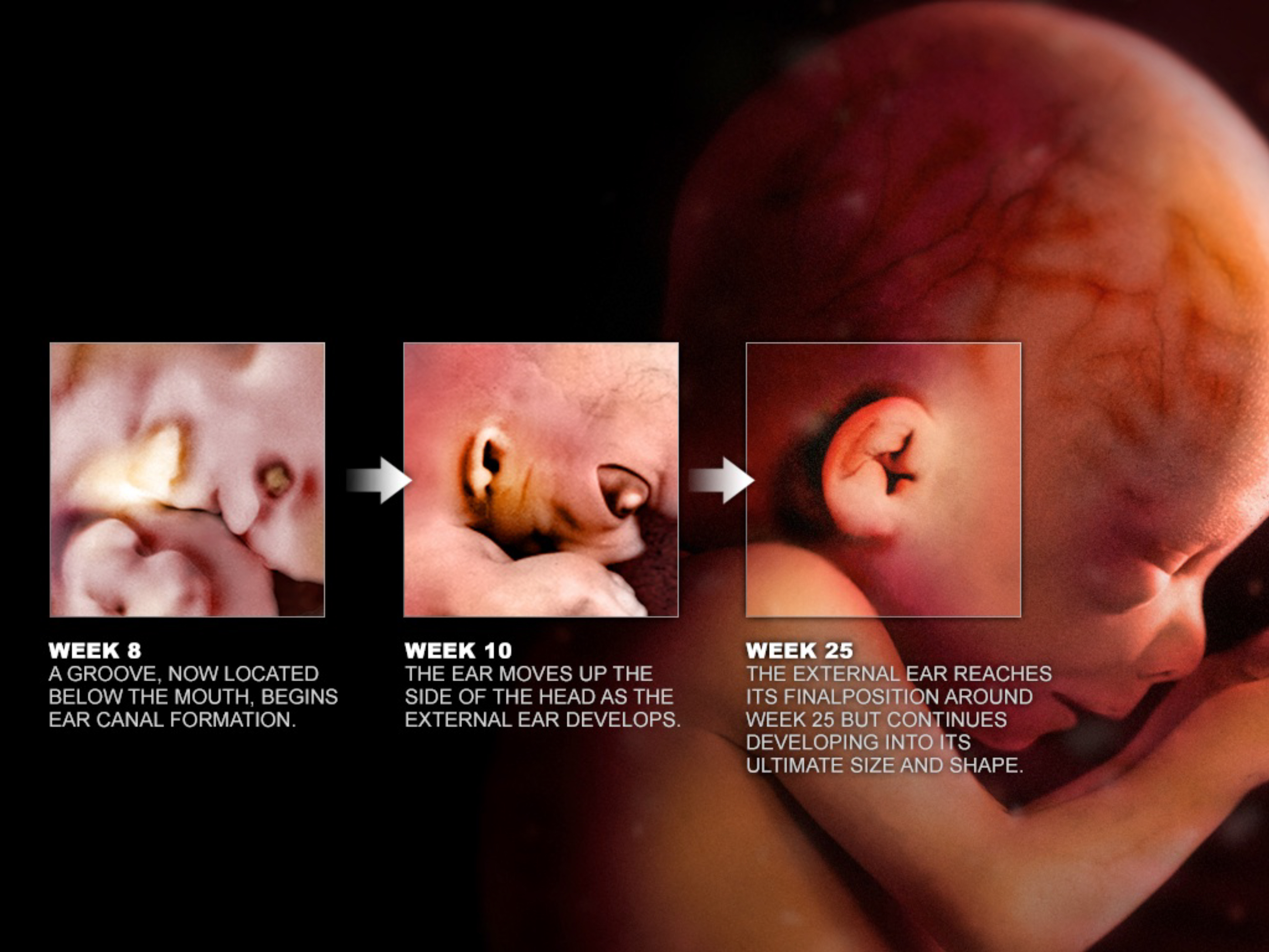
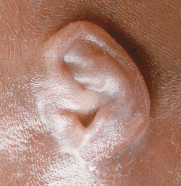
By just 22 days after conception, or five weeks gestation, the unborn child’s ear has already begun forming.1 2 The inner ear, responsible for detecting sound and motion, starts forming first. The outer ear – the visible shell-shaped tissue responsible for collecting and localizing sound, starts forming next. The middle ear, responsible for amplifying sound and stabilizing air pressure in the ear, forms from the interactions of surrounding tissues. Over the following weeks, these parts continue to grow and mature, setting the foundation for hearing.

(Image Credit: Science Source)
How does the inner ear form?

The inner ear is the first to form. In week 5, a tiny patch of surface tissue near the future brain called the otic placode thickens and sinks inward to form the otic pit. Between 28 and 33 days post conception, or 6 weeks gestation, the sides of the otic pit fuse to create a bubble of tissue called the otic vesicle. By week 7, the otic vesicle forms two regions. The frontal section forms the cochlea, which allows a person to hear, while the section closer to the back becomes the vestibular organs, which help a person balance and sense motion.3 4 In week 8, the back portion of the otic vesicle extends outward to form the vestibular organs.5 Also in week 8, the frontal portion of the otic vesicle forms a long tube – the early cochlea. It grows in a spiral like a snail shell so that by week 12 it has completed 2 ½ turns.6 Between 12 and 14 weeks, the hair cells within the cochlea, which turn sounds into neural signals, start growing.7 By 18 weeks, the bones protecting the inner ear start hardening from cartilage.8 All of the neural structures underlying hearing are in place by 22 weeks gestation and the inner ear has reached its adult size.9 10

How does the middle ear form?

The middle ear includes the eardrum – bones for amplifying sounds, and the eustachian tube. The bones of the middle ear start forming around week 8 from the first and second pharyngeal arches.11 Pharyngeal arches are paired bulges of tissue on either side of the embryo’s neck. The bones of the middle ear start as cartilage, then slowly harden to become bone. As the fetus develops, the tissue surrounding these bones breaks down to create an air-filled tympanic cavity with the ossicles hanging inside.12

The Eustachian tube is a thin passage connecting the middle ear to the back of the nose and throat to help drain fluids and equalize air pressure. The Eustachian tube starts forming in week 7 by elongating the first pharyngeal pouch, which is a pocket of tissue near the developing throat. This passage grows rapidly between 18 and 30 weeks gestation. At birth, the eustachian tube is shorter, narrower, and more horizontal than in later childhood, which makes it more difficult to drain fluids. That is why infants have more ear infections than older children.13

How does the outer ear form?

Around 6 weeks gestation, the outer ear starts as a groove on the neck, just below the mouth. Over the next two weeks, 6 bumps called the auricular hillocks form and fuse together to create a shell shape.14 The outer ear also migrates towards its final position on the side of the head.15 From there, the outer ear forms more folds, reaching a mature adult shape around 24 weeks.16 The ear canal is open by 20 weeks,17 but relatively short. The ear canal does not reach its adult length until about age 9.18

The outer ear’s shape helps a person localize sounds; and since every ear shape is slightly different, everyone’s brain has made slightly different connections so that people have trouble telling the direction of sounds that are recorded inside someone else’s ear.19 Oddly enough, the outer ear actually continues to grow for a person’s entire life.20

The ear at four months gestation. (Image Credit: <a href="https://www.priestsforlife.org/graphic-images/index.aspx?gid=1&sid=1">Priests for Life</a>)
The ear at four months gestation. (Image Credit: Priests for Life)
When does the fetus start hearing?

Scientists studying fetal hearing have found that the auditory system begins responding to sound surprisingly early in pregnancy. In one experiment, a 19-week fetus moved after exposure to a loud, low-frequency tone, like a car horn.21 Younger fetuses tend to require louder sounds to provoke a reaction and appear more sensitive to lower-pitched tones with frequencies similar to those found in adult male voices.22 As with many developmental milestones, the timing varies. By 20–21 weeks, about half of fetuses show detectable changes in heart rate when exposed to sound.23


In one experiment, a 19-week fetus moved after exposure to a loud, low-frequency tone, like a car horn.24

Responses become more consistent as the auditory system matures. Around 24 to 25 weeks, many fetuses show startle responses or body movements when exposed to tones comparable to a lawn mower.25 26 By 28 weeks, heart-rate accelerations in response to sound are consistently observed,27 28 and brain recordings indicate that fetuses can even distinguish between tones of different pitch.29 Between months 6 and 7, the unborn child consistently responds to external sounds, showing awareness of the world beyond the womb.