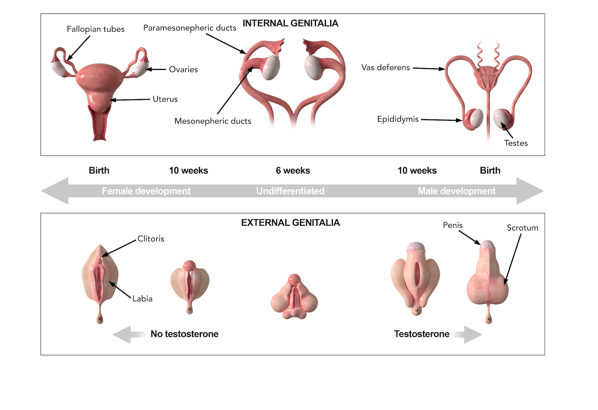
close-panel

Charlotte Lozier Institute

Phone: 202-223-8073
Fax: 571-312-0544

2776 S. Arlington Mill Dr.
#803
Arlington, VA 22206

Get Notifications

Sign up to receive email updates from Charlotte Lozier Institute.

Male vs. Female Development

Dive Deeper

A person’s sex is determined at conception. Males have a set of XY chromosomes and females have a set of XX chromosomes. These sex chromosomes determine whether the embryo develops as a male or female. Initially the gonads in both male and female embryos can develop as either testes or ovaries.  Starting at 8 weeks gestation, an embryo with a Y-chromosome produces a factor known as SRY that simultaneously causes the gonads to begin maturing as testes and prevents them from maturing as ovaries.  The testes, in turn, produce two important hormones:  testosterone and anti-Mullerian Hormone, or AMH. In males, the testes starts producing and releasing testosterone by 10 weeks. Testosterone allows the primitive male anatomy to mature, while AMH causes the primitive female structures to shrivel. The male internal ducts consist of a pair of tubes called the mesonephric ducts that will mature into the vas deferens and epididymis. The epididymis stores sperm, and the vas deferens is the sperm duct.  1

At 10 weeks gestation, the external genitalia still look the same between boys and girls, but over the next 4 weeks they will become very distinct. Illustration: Reproductive organ development (Image Credit: Science Source)

In females, a different signaling molecule causes the bipotential gonads to develop into ovaries and prevents them from developing into testes. The female structures also consist of a pair of tubes called the paramesonephric ducts. Parts of the paramesonephric ducts will later fuse at the midline to form the uterus, cervix and upper portions of the vagina, while the unfused sections becoming the fallopian tubes. In the absence of testosterone and anti-Mullerian Hormone, the primitive female anatomy matures, and the male structures, that require testosterone to survive, starts to shrivel. Therefore, by default, a fetus will develop as a female unless genes from the Y chromosome activate the correct signaling pathways to start male reproductive development.2

Externally, both male and female embryos have similar genitalia for the first seven weeks gestation.3  Because of this, early ultrasounds cannot typically determine whether the baby is a boy or a girl.  Both sexes develop folds of tissue adjacent to the external openings that lead to the bladder and the intestines. Both sexes also have a small protrusion of tissue known as the genital tubercle. Prenatal DNA testing can determine early on whether a baby is male or female before being able to detect anatomical differences by ultrasound.

Illustration: Sexual differentiation of the genital duct system (Image Credit: NIH National Library of Medicine)

Around 12 weeks gestation, the external structures start to differentiate.  In males, the genital tubercle elongates and becomes the penis. The external folds fuse at the midline and become the scrotum. By 4 months, the male genitalia are mostly formed. In females, the genital tubercle elongates only slightly, forming the clitoris, and the surrounding folds become the labia.  Most of the female reproductive structures are correctly positioned by 14 weeks.  At this point, a doctor can usually tell if the baby is a boy or a girl by using an ultrasound.4

Sperm-egg fusion