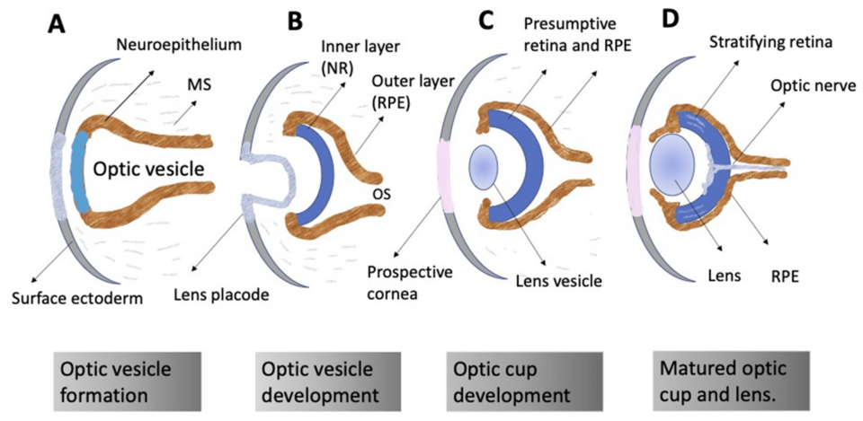
How do photoreceptors form?
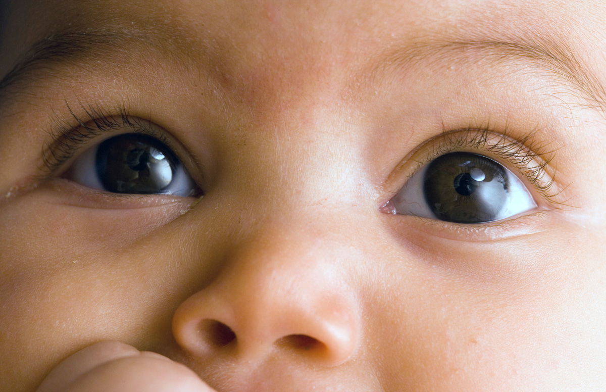
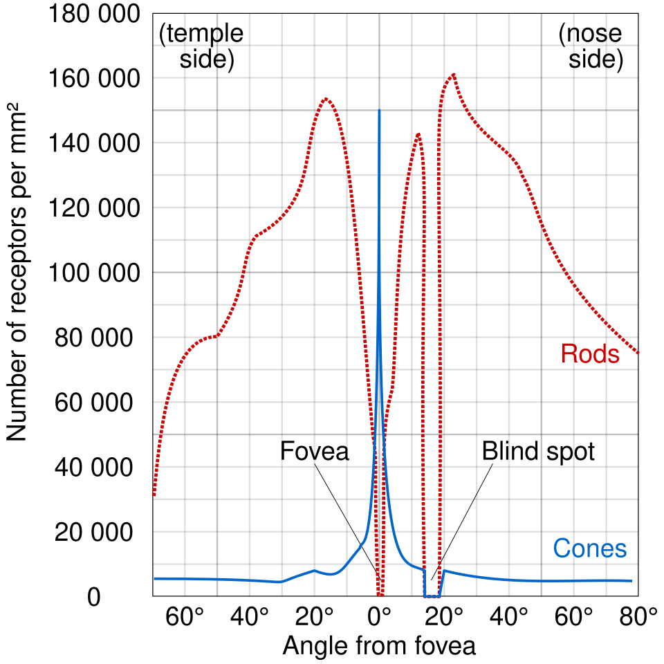
The retina, located at the back of the eye, contains two types of light-sensitive cells called rods and cones. These cells begin forming around 11 weeks.19 Rods are extremely sensitive – capable of detecting a single photon—and are responsible for seeing in dim light, while cones function best in bright light and enable color vision. In adults, each eye typically contains about 100 million rods and 6 million cones.20 While both rods and cones are formed all over the retina, cones cluster in the center, called the fovea, and rods are concentrated in the surrounding area, called the periphery.21
The organization of the retina continues to change throughout pregnancy and even after birth. Cell division in the fovea stops around 16 weeks of gestation, while cell division in the outer parts of the retina continues until about 32 weeks.22 As development proceeds, cones gradually migrate toward the center of the retina, while rods move outward—a process that continues for many months postnatally.23 Between 18 and 30 weeks, the retina overproduces relay neurons known as ganglion cells, resulting in a temporary surplus compared to the number present at birth.24
Despite their early appearance, rods are not yet fully functional; in premature infants born around 36–38 weeks, the retinal response to light is only about 2% as strong as that of an adult retina.25 Similarly, the cones continue to develop well after birth, reaching maturity around four years of age.26


.png)