The
Voyage of Life

Women Have Real Choices: Did you know? There are 15 better women’s service providers for every Planned Parenthood center.

Abortion Drug Facts: Research shows the rate of abortion pill-related emergency room visits has increased.

Pregnancy Center Reports: Read about the work of pregnancy centers nationally and at the state level.

Pro-Life Safety Net Reports: State-run programs in pro-life states that serve families, fathers, pregnant women and unborn children.
Evaluation Of Human Fetal Tissue Research: Learn about federal funding for research using human fetal tissue.

Stem Cell Research Facts: Learn about the incredible possibilities of stem cell research.

Signs of Life: One of the most striking things about the realm of science fiction is that it is seldom fiction.

State Abortion Reporting: Learn about state abortion reporting.

Facts not Fear: Will Pro-Life State Laws Hurt Women and Hinder Doctors?

Assault on Science: Refuse to let journals and editors launch meritless attacks without a fight.
Women Have Real Choices: Did you know? There are 15 better women’s service providers for every Planned Parenthood center.
Abortion Drug Facts: Research shows the rate of abortion pill-related emergency room visits has increased.
Pregnancy Center Reports: Read about the work of pregnancy centers nationally and at the state level.
Pro-Life Safety Net Reports: State-run programs in pro-life states that serve families, fathers, pregnant women and unborn children.
Evaluation Of Human Fetal Tissue Research: Learn about federal funding for research using human fetal tissue.
Stem Cell Research Facts: Learn about the incredible possibilities of stem cell research.
Signs of Life: One of the most striking things about the realm of science fiction is that it is seldom fiction.
State Abortion Reporting: Learn about state abortion reporting.
Facts not Fear: Will Pro-Life State Laws Hurt Women and Hinder Doctors?
Assault on Science: Refuse to let journals and editors launch meritless attacks without a fight.
Your donation helps us continue to provide world-class research in defense of life.
DONATEPhone: 202-223-8073
Fax: 571-312-0544
2776 S. Arlington Mill Dr.
#803
Arlington, VA 22206
Heartbeat detectable by ultrasound
The heart keeps beating rhythmically, moving oxygenated blood throughout the unborn baby. This week the heartbeat can be consistently detected by ultrasound.4 Clusters of cells start growing in just the right locations to form the upper and lower limb buds. These buds will become the arms and legs.5
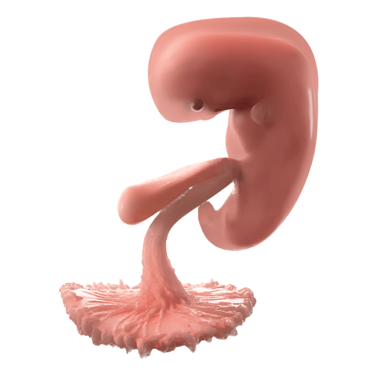
![This composite image rendered from light-guided endoscopy recordings shows an embryo alive in the womb at 4 weeks following [tooltip anchor="fertilization"]Sperm-egg fusion[/tooltip] (or 6 weeks gestation). Note how the arm and leg buds have formed, and the heart is clearly visible within the chest. (Image Credit: <a href="https://erf.science/#high-resolution">Education Resource Fund</a>)](https://lozierinstitute.org/wp-content/uploads/2021/08/IMG_04-ERF-4-Week-Embryo_FULL-Large.jpeg)
Last week, at only 22 days after conception or 5 weeks and 1 day gestation, the embryo’s heart started beating. At 6 weeks gestation the unborn baby has a steady heartbeat around 110 beats per minute!6 In week 6, the heartbeat can consistently be detected by ultrasound in viable pregnancies.7
The heart moves oxygen-carrying blood throughout the unborn baby so that he can continue to grow. Without an early heartbeat that circulates blood, new tissues would not have enough oxygen to survive.8 Once the heart starts rhythmically beating, it will not stop until the person dies.
The baby’s heart beats much faster than the mother’s, with a heartrate ranging from 95 to 149 beats per minute. The unborn child’s heartrate peaks during gestational week 9 at over 170 beats per minute.9 The heart beats about 54 million times between conception and birth.10
Yes! Clinicians use ultrasound to measure the rhythmic movement of the beating heart to determine the heartrate and health of the baby. Ultrasound does not measure electrical activity; it measures pulses of high-frequency sound reflected off solid objects, like the heart pumping blood. A doppler ultrasound can also measure motion by detecting changes in the frequency of the sound as it bounces off moving heart tissues and blood to accurately measure heart rate.12 13 Just as a cell phone camera uses light bouncing off an object to take its picture, the ultrasound equipment uses sound bouncing off the early heart to create the image and sounds that a mother sees and hears on the screen. Using reflected sound to measure the heartbeat instead of light does not make the heartbeat any less real—instead it is scientific proof that the baby’s heart is real!14
While it may seem that these dates are incongruent, both are scientifically accurate because they arise from two separate dating systems. Embryologists determine the age of the unborn based on fertilization, the beginning of human life. Most obstetricians, medical professionals, and mothers use gestational age based on the mother’s last menstrual period. Gestational age is typically two weeks greater than embryonic age, because a woman usually ovulates two weeks after her last menstrual period.
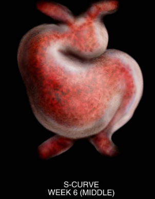
As the week progresses, the heart tube increases dramatically in length by adding new heart muscle cells.15 As the heart tube grows, it starts to bend to the right, and different portions expand. The motion and pressure of the blood pumping within the heart causes the heart cells to change their growth patterns to create the bend. The heart tube will become the left ventricle and left and right atrium, while the right ventricle forms from nearby cells in the secondary heart field. Cells from the secondary heart field will also help establish the major veins that lead into the mature heart.16
The face develops from the first pharyngeal arch and neural crest cells. Pharyngeal arches are paired bulges of tissue on either side of the embryonic neck. Each one has its own blood vessel, nerve, and cartilage, which will later turn into bones. Around week 6, the unborn baby grows six pharyngeal arches in the head and neck area. Neural crest cells are special cells that grow near the neural tube and play an important role in forming the face.17
Also around week 6, the face begins to form from five swellings of tissue from the first pharyngeal arch
and neural crest cells. These facial prominences include:

![This baby's eye began forming 22 days after [tooltip anchor="conception"]Sperm-egg fusion[/tooltip]. (Image Credit: Adobe Stock Photos)](https://lozierinstitute.org/wp-content/uploads/2021/08/AdobeStock_14621026.jpeg)
Last week, the top and bottom of the neural tube sealed shut, creating the brain and spinal cord. The eye also began to form last week, 22 days after conception.20 This week, pockets of neural tissue grow from the young brain towards the surface of the face. Those pockets of neural tissue, also known as the optic vesicles, will become the optic nerve, retina and other structures in the eye. The optic vesicles cause some of the surface tissue to separate, thicken, and start forming the lens of the eye.21 Later, the lens will become transparent. This occurs as lens cells lose their organelles and develop special orderly crystalline proteins.22 Most of the lens’s crystalline proteins form around birth. The lens grows rapidly in the womb and for the first year of life, then more slowly thereafter so that most of the lens fibers in an elderly person have been there since before birth.23
By the start of week 6, a simple digestive tube runs from the unborn baby’s head to rump.24 Near the top, the esophagus forms and elongates quickly.25 Just below, the digestive tube enlarges and rotates, gradually becoming the stomach.26 At the same time, the liver and pancreas begin to grow, preparing to regulate nutrients in the baby’s body.27 By week 8, the pancreas fuses into one organ.28 Around 10 weeks, the gut starts producing hormones like insulin.29 Though the baby won’t eat until after birth, the digestive system starts practicing as the unborn baby swallows amniotic fluid, starting around 12 to 13 weeks gestation.30 31 32 From the very beginning, the unborn baby develops not as a collection of unrelated parts, but as a coordinated human body growing toward maturity.
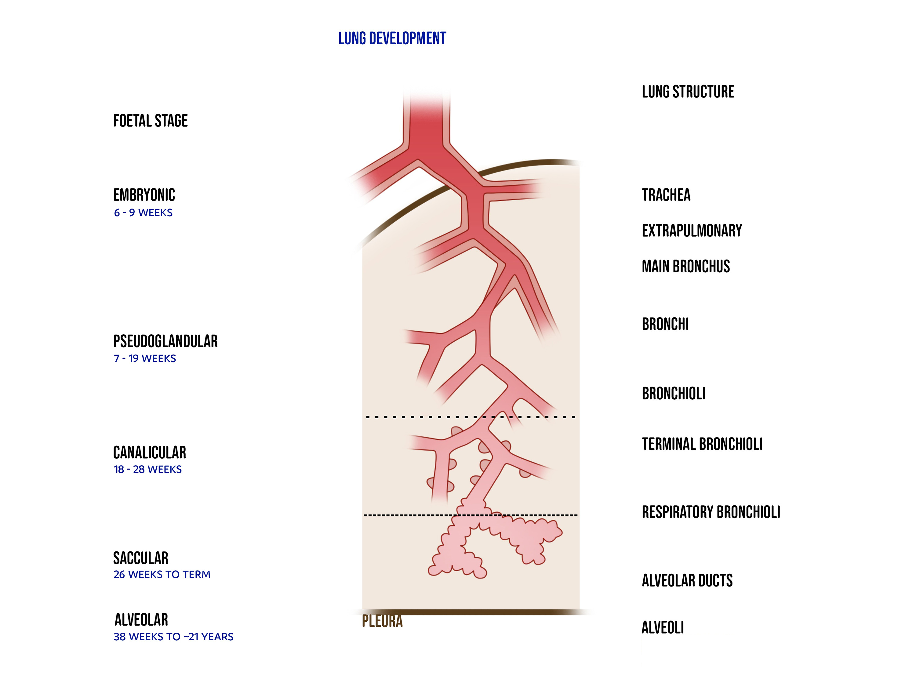
Unlike many other vital organs, the baby does not need the lungs to function in the womb, since the baby receives his oxygen from the placenta. Nevertheless, lung development begins early. Between 5 and 6 weeks, two small lung buds form and connect to the developing airway. By 7 weeks, these buds branch into the main bronchi, which further divide into the lobes of the lungs. Over the next 12 weeks or so, these airways branch about twenty times, each time forming smaller tubes called bronchioles. Blood vessels grow alongside them, preparing for future oxygen exchange, even though the baby does not yet breathe air.33
Between 18 weeks and birth, the lungs begin forming tiny air sacs, where oxygen and carbon dioxide exchange will eventually occur. By about 22 weeks, the lungs start producing surfactant, a slippery substance that keeps these air sacs open. Surfactant is pivotal for survival outside the womb.34 Between 29 weeks and birth, the lungs expand over four times in size to prepare for breathing.35 At birth, the baby will have developed 150 million alveoli, about half of that seen in adults.36 After birth, lung growth continues dramatically: most mature alveoli form after delivery, dramatically increasing the surface area needed for gas exchange.37 Most of this growth happens in the first six months,38 but continues through childhood and even into early adulthood—up to around 21 years of age.39
![This medical visualization was created from human scanned data. The nervous system develops from the neural tube, which begins to form around the 22nd day after [tooltip anchor="conception"]Sperm-egg fusion[/tooltip]. Once the neural tube closes around the 28th day after conception, specific regions and structures in the brain begin to form. (Image Credit: Science Source)](https://lozierinstitute.org/wp-content/uploads/2021/08/33daybrain_ScienceSource.png)
By 6 weeks gestation, the baby’s head appears strikingly large due to the brain’s rapid growth.40 Inside, the early brain is dividing into five major regions. The front portion will expand into the cerebral hemispheres, the center for thinking, decision-making, and perception. Just behind it, the thalamus begins forming, a crucial relay station that will route sensory information through the brain. Farther back lies the midbrain, which helps locate moving objects and sounds. The back of the brain develops into the cerebellum and brainstem, structures essential for movement, balance, and basic life functions. At this stage each region resembles a small bubble of tissue, with the back of the brain continuous with the developing spinal cord.41 Protective membranes called the meninges also start developing.42
This early window of development is critical. Between about 21 and 28 days after conception, the neural tube—the structure that will become the brain and spinal cord—must close properly. If it does not, serious conditions such as spina bifida or anencephaly can occur.43 Because this process happens before many women realize they are pregnant, public-health efforts focus on prevention. In 1998, the United States began fortifying bread and cereal products with folic acid, a vitamin essential for neural tube formation.44 The policy has had a measurable impact: researchers estimate that folic acid fortification prevents roughly 1,300 cases of spina bifida each year.45
The aorta is the biggest blood vessel in the human body. It’s like a superhighway for blood, carrying it from the heart to the rest of the body. But how does this huge vessel form?
Early in week 6, the aorta starts as two parallel tubes with six pairs of arches arranged like delicate loops on either side of the early throat region. These arches form in a specific sequence and begin to transform rapidly. Some arches disappear. Others enlarge. The ones that remain twist, rotate, and fuse with remarkable precision creating the asymmetrical aortic arch. The aorta is the blood vessel that will carry oxygen-rich blood from the tiny, beating heart to the rest of the growing body.46 All of this occurs while the baby’s heart rate averages from 100 to 134 beats per minute!47
By the end of week 8, the final configuration of the aorta begins to emerge. It curves upward from the heart, then loops around down the spine, ready to serve the lungs, brain, and every developing organ. The aorta becomes the main pipeline for life-giving blood.48