Search Icon
close-panel

Charlotte Lozier Institute

Phone: 202-223-8073
Fax: 571-312-0544

2776 S. Arlington Mill Dr.
#803
Arlington, VA 22206

Get Notifications

Sign up to receive email updates from Charlotte Lozier Institute.

This field is for validation purposes and should be left unchanged.

close-panel

Become A Defender of Life

Your donation helps us continue to provide world-class research in defense of life.

DONATE

Charlotte Lozier Institute

Phone: 202-223-8073
Fax: 571-312-0544

2776 S. Arlington Mill Dr.
#803
Arlington, VA 22206

Week 8

Brain activity and new movement patterns

Human Prenatal Age
  • Post-conception week 6
  • Days of life 42-48
  • Gestational Week 8
Highlights
  • The unborn baby can quickly move his arms or legs, as if startled.1

  • The unborn baby’s brain is already active—neurons are connecting and producing early brain waves.2

  • The unborn baby’s face is forming, with eyes, a nose, a mouth, and even a tiny tongue.3

Brain cells, called neurons, have already connected to one another. Networks of neurons produce immature patterns of brain activity, or “brain waves,” that have been recorded during surgeries on ectopic pregnancies when the embryo was the size of a jellybean at 8 ½ weeks gestation.4

This composite image rendered from light-guided endoscopy recordings shows the embryo alive in the uterus 6 weeks after [tooltip anchor="fertilization"]Sperm-egg fusion[/tooltip] (or 8 weeks gestation). Notice how the hands and feet are forming. This unborn baby would move away from a light touch near his mouth. (Image Credit: <a href="https://erf.science/#high-resolution">Education Resource Fund</a>)
This composite image rendered from light-guided endoscopy recordings shows the embryo alive in the uterus 6 weeks after fertilization (or 8 weeks gestation). Notice how the hands and feet are forming. This unborn baby would move away from a light touch near his mouth. (Image Credit: Education Resource Fund)
Embryonic brain activity at 8 weeks of pregnancy

At 8 weeks gestation, neurons start moving away from their birthplace in the brain to form the interconnected and layered cortex.5 Measurable brain activity has been recorded in human embryos as early as 45 days after conception, or 8 weeks and 3 days gestation, showing rhythmic electrical patterns rather than random neural firing.6 These early EEG signals suggest that neurons are already communicating and synchronizing their activity, even displaying patterns similar to sleep states seen later in development.7 As pregnancy progresses, brain activity becomes more complex, eventually allowing researchers to detect responses to light, sound, and other stimuli in the womb.

Neurons and muscles are connected as well. In week 8, the unborn baby can quickly move his arms or legs, as if startled.8 The unborn baby also moves away from a light touch to the nose or chin.9

Measurable brain activity has been recorded in unborn babies as early as 45 days after conception.10
Making muscles

Skeletal muscles allow people to move their limbs and hold themselves upright. They are responsible for the first fetal movements observed at 7 ½ weeks.11 But how do they form?

Muscles arise from special clusters of cells called somites, which line up neatly along the developing spine. Inside each somite, certain cells receive chemical signals that tell them to become muscle-forming cells, or myoblasts. The myoblasts multiply and fuse together, forming long tube-like fibers that become muscle tissue. As the early fibers bundle together and get wrapped in connective tissue, they become the muscles that power every movement in the body.12

Even after birth, the muscles keep growing. In fact, every time a person exercises or recovers from injury, a team of satellite cells — a form of stem cells — help repair the muscle tissue. Satellite cells can even grow new muscle fibers.13

The human heart at 8 weeks gestation. This week the heart divides into a left and right side.[citation text="Buijtendijk, M. F., Barnett, P., & Van Den Hoff, M. J. (2020, March). Development of the human heart. In American Journal of Medical Genetics Part C: Seminars in Medical Genetics (Vol. 184, No. 1, pp. 7-22). Hoboken, USA: John Wiley & Sons, Inc... https://onlinelibrary.wiley.com/doi/full/10.1002/ajmg.c.31778." href="https://onlinelibrary.wiley.com/doi/full/10.1002/ajmg.c.31778"] (Image credit: Science Source)
The human heart at 8 weeks gestation. This week the heart divides into a left and right side.14 (Image credit: Science Source)
Heart development at 8 weeks gestation

As the unborn baby’s heart continues to grow at 8 weeks of pregnancy, it undergoes an extraordinary transformation into its familiar four-chambered shape. Thin walls called septa start dividing the heart into right and left sides. These walls develop from a special, jelly-like material that gives early structure to the heart. Meanwhile, the ventricles—the lower pumping chambers—grow rapidly, as new cells are added and muscle tissue thickens to prepare for a lifetime of work.15 One study in mice found that ventricles might increase in size nearly 100-fold!16

Though the atria, the upper blood-collecting chambers, appear symmetrical at first, each plays a unique role from the beginning. The right atrium absorbs early veins feeding from the body into the heart while the left atrium absorbs the pulmonary vein, which will carry oxygenated blood from the lungs after birth.17 These side-specific changes happen in a precise sequence to create a healthy and functional heart.

How does the ear form?

By week five, ear development begins. A small patch of surface tissue near the future brain called the otic placode starts to thicken and sink inward. This forms the inner ear, which will help with hearing and balance.18 19

In week 6, the otic placode forms a pouch called the otic vesicle.20 In week 7 the otic vesicle grows longer and by week 8, it twists and folds to make parts of the inner ear, including the early cochlea— which helps with hearing — and the semicircular canals —which help with balance.21 22 The early cochlea grows in a spiral fashion so that by week 12 it has completed 2 ½ turns.23 At the same time, inside the cochlea, the structures needed to turn sound waves into neural signals start forming.24 The inner ear reaches its adult size and shape around 22 weeks.25 26

Making the Middle Ear

Around week 8, a new part of the ear begins to form from a pouch in the neck.27 This turns into the middle ear, which includes the eardrum and three tiny bones — the smallest bones in the body — that amplify sound. The Eustachian tube, a narrow tube that connects the baby’s middle ear to the back of his nose and throat, also starts to grow.28

Forming the Outer Ear

Starting in week 6, little bumps on the side of the head start to develop into the outer ear, or the part visible on either side of the head. These bumps move towards their final position and establish their shell shape around week 24.29

Fine-Tuning and Connecting

Between 7 and 20 weeks, all these parts connect.30 31 Tiny hairs grow inside the cochlea to detect sound waves. Every hair cell in the cochlea is specially tuned for a specific pitch. Most of the tuning of hair cells occurs between 28 weeks gestation and the first few months outside the womb.32

In summary, the ears start forming when the baby is just a few weeks old in the womb—and by about 24 weeks gestation, the inner ear can detect sounds outside the womb!33 34
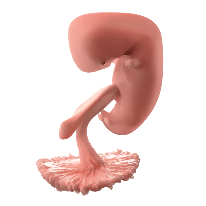
The development of the outer ear from gestational week 8 (6 weeks after [tooltip anchor="conception"]Sperm-egg fusion[/tooltip]) to month 5.(Compilation image credit: Katrina Furth, Ph.D. using images from <a href="https://erf.science/#high-resolution">Education Resource Fund</a>)
The development of the outer ear from gestational week 8 (6 weeks after conception) to month 5.(Compilation image credit: Katrina Furth, Ph.D. using images from Education Resource Fund)
By week 7, the first structures for smell are forming—laying the foundation for millions of scent-detecting cells in the unborn baby’s nose. (Image Credit: Adobe Stock Photos)
How does the nose develop?

By the end of week 6 of pregnancy, two small areas of thickened skin appear on each side of the bulge of facial tissue called the frontonasal prominence. These areas are called nasal placodes, and they grow larger as the baby develops. During week 7, the middle of each nasal placode folds inwards to form depressions called nasal pits. These nasal pits eventually develop into the olfactory epithelium, the specialized tissue that makes up the inside of the nose and allows people to smell. The olfactory epithelium gives rise to olfactory receptor neurons, which start sending their axons towards the developing brain in week 8.35 Also in week 8, the nasal cavity starts to connect to the back of the throat.36

Folds of tissue surround the nasal pits to form the nostrils, in which human beings have millions of olfactory sensory neurons distributed along the surface!37 38 By 11 weeks gestation, the nose has developed its recognizable shape.39 The nostrils remain plugged with thin tissue from 9 weeks to about 18 weeks’ gestation,40 41 but amniotic fluid still enters the nasal cavities when the baby starts swallowing around gestational week 12 .42

How does the unborn baby's liver form?

In the middle of week 5, the liver begins forming as a small bud emerging from the unborn child’s developing digestive system.43 Like a carefully spun web, finger-like projections stretch and weave together to form stringy cords. Different types of cells nestle among the cords to create the liver. Oxygen-rich blood from the umbilical cord flows directly through this developing organ, creating its shape and bright red color. The blood flow determines the size of the liver’s lobes, with the right side growing larger than the left.44 By week 8, this growth happens so quickly that the fetal intestines temporarily grow outside the body into the umbilical cord, only to return a few weeks later around week 12.45 46 Even in these earliest stages, the unborn child’s body is growing, changing, and thriving — a powerful witness to the humanity and dignity of life before birth.

The unborn child’s liver performs incredible tasks from an early age. In adults, the liver filters blood coming from the digestive system, produces bile, and helps regulate blood sugar levels. But in the unborn baby, the liver takes on another deeply vital responsibility: producing blood. Between weeks 5 and 7, blood-producing cells move into the unborn baby’s liver. By 8 weeks, the liver becomes the primary site of blood formation. In fact, during weeks 13 and 14, almost 60% of the cells in the liver make blood.47 For nearly five months, the liver is the main source of both red and white blood cells until around the 7 months of pregnancy, when the bone marrow takes over.48 49 The liver stops making blood cells about 1-2 weeks after birth.50

The liver creates most of the baby's red blood cells from 2 to 7 months of pregnancy. (Image Credit: <a href="https://commons.wikimedia.org/wiki/File:Red_Blood_Cell.jpg">Wikimedia commons</a>)
The liver creates most of the baby's red blood cells from 2 to 7 months of pregnancy. (Image Credit: Wikimedia commons)
Continued development at 8 weeks of pregnancy

At 8 weeks gestation, in the second month, the baby’s face, head and neck continue developing. Eyelids start forming,51 and pigments in the retina make the eyes more visible.52 Salivary glands start developing inside the mouth.53 By the end of this week, key breathing and crying structures—the voicebox, trachea, and early lungs—have formed.54

At the same time, the baby grows quickly—about 1 millimeter each day.55 56 Growth happens in bursts, especially in the arms, legs, and head. The hands develop one or two days before the feet. Tiny ridges called digital rays appear in the hands and feet, marking where fingers and toes will form.57


Dive Deeper
The outer ear becomes shell-shaped around ten weeks gestation...
The earliest brain activity was recorded from a 45-day-old embryo...
Sperm-egg fusion
Sperm-egg fusion
Electroencephalogram for measuring electrical activity of the brain
Little patch destined to become the inner ear
Sperm-egg fusion