


Q&A with the Scholars: Fetal Brain Development and Pain-Capability


Katrina Furth, Ph.D., earned her doctorate in neuroscience at Boston University, performing her dissertation research at the National Institutes of Health. Since graduating, she has worked as an adjunct professor at Marymount University in Arlington, Virginia. Dr. Furth enjoys educating scientists and non-scientists alike about brain development and leads seminars and talks about the neuroscience of the unborn. In this interview she discusses fetal neurological development.

Katrina Furth, Ph.D.
According to the Centers for Disease Control and Prevention (CDC), in 2014, 91.5 percent of the abortions reported by gestational age – more than 400,000 of the 440,330 for which gestational age is known – were performed at or before 13 weeks of gestation. Can you tell us about the development of the nervous system of an unborn baby at this time, roughly 11 weeks old since conception? Along those same lines, can you explain how much of a baby’s nervous system has formed by the time a woman even realizes that she is pregnant?
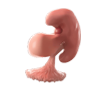
Katrina Furth: A woman will not realize that she is pregnant until she has first missed her period, which, depending on her history of menstruation, will be 2-4 weeks after conception (4-6 weeks gestation). Many important systems develop during this time. One week after conception, the embryo implants in the uterus. Following implantation, the embryo establishes different sides and cell layers based on chemical gradients from the cells that surround it. The neural tube, which becomes the brain and spinal cord, seals up completely three weeks after conception, one day after the fetal heart begins to beat. By the fourth post-conception week, the eyes and ears have started to form, including connections to the developing brain.
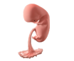
Ultrasounds may detect the first fetal movements as early as five weeks post-conception. These movements provide evidence of electrical impulses at the connection between nerves and muscles. In the sixth week, the forebrain – the brain region responsible for processing perception, thoughts, and decisions in adults – doubles in size. In fact, the brain starts growing at a rate of 250,000 neurons per minute for the next 21 weeks! In the seventh week, the fetus starts showing a preference for his right or left hand. By 11 weeks post-conception, the fetus performs complex behaviors that require working neural circuits, including hiccupping, stretching, grasping objects, and turning away from loud noises.
Importantly, the formation of the human brain dominates early development. In fact, at eight weeks post-conception, the fetal brain weighs 43 percent of its total body weight. By comparison, a newborn’s brain weighs 10 percent of his total body weight, and an adult’s brain weighs just 2 percent of his total body weight. Therefore, even early fetal development occurs in connection with neural activity.
How do we know that a six-week-old post-conception human fetus has brain activity?
Katrina Furth:Typically, doctors record brain activity, or EEGs, using electrodes placed on the scalp. However, most fetal EEGs are recorded during labor, or in the third trimester by placing a huge array of electrodes all over a mother’s abdomen. Neither technique would work for a six-week-old fetus, who is about the size of a grape.
In 1955, Winslow Borkowski and Richard Bernstine, doctors at Jefferson Medical College Hospital, took advantage of some tragic medical emergencies. These doctors temporarily preserved two tiny unborn children removed during ectopic pregnancies at 45 days post conception and 77 days post conception. These doctors recorded from these fetal brains and observed patterns of brain activity also seen later in development. The team inserted needle electrodes to record brain activity below the brain surface. These electrodes penetrated the cranial tissue, which solves the problem of electrical interference from nearby muscles. These studies ended in the 1960s.
When can a human fetus feel pain, and what methods are used to determine this?
Katrina Furth: Importantly, whether a fetus can feel pain has no bearing on the fact that the human is a unique individual worthy of protection from the first moment of his or her conception.
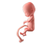
The best evidence suggests that the fetus starts reacting to pain around 7.5 weeks post-conception because the fetus consistently moves away from painful stimuli in ultrasound recordings. Furthermore, by examining the skin of aborted fetuses, scientists have found that pain receptors cover fetal skin by nine weeks post-conception, with the largest concentrations of pain receptors clustered in the fingers and face.
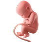
A major debate centers on the question of whether a fetus perceives pain before a two-step neural pain circuit has formed. Pro-choice lawyers and doctors have argued that the fetus cannot perceive pain until connections exist between the pain receptors in the skin and the thalamus, and connections exist between the thalamus and the cortex. Unfortunately, it is difficult to prove the existence of these connections in living fetuses without hurting the fetus; scientists only consistently observe these connections from outside the womb at 21 weeks post-conception, although they are observed a month earlier in aborted fetuses. Importantly, fetuses show hormonal stress responses to pain during prenatal surgeries performed at 16 post-conception weeks, strongly suggesting a complete pain perception pathway has formed by this time.
However, I would also argue that the connection between the thalamus and the cortex is less important for pain perception than the pain receptors themselves. First, pain responses are sometimes intact in adults in an unresponsive wakeful state (referred to by some as a “vegetative” state). Second, patients with rare congenital diseases that cause them not to feel pain, have intact cortico-thalamic connections, but abnormal pain receptors. Together, this evidence suggests that pain receptors are both necessary and sufficient to produce pain.
Some abortion advocates use partial neurological development as an argument for the moral legitimacy of abortion. Can you tell us what you think about this argument, and explain when the brain is fully finished developing?
Katrina Furth: Partial neurological development has no bearing on whether a human has a right to life and protection. Unless we are willing to accept the murder of premature babies, infants, children, and adolescents as ethical, I believe that we should not accept any arguments based on the idea of partial neurological development.
Importantly, brain development continues long after birth. In fact, the final step in brain development, myelination, does not finish until around age 25. Given the near-universal agreement on the protection of newborns and children, arguments based on partial brain development fall apart.
Interestingly, newborns are born with 10-40 percent more neurons than they will need as adults, and two-year-olds have twice as many synaptic connections between neurons than they will need as adults. The next decade is spent refining all of these synaptic connections so that one neuron innervates one muscle, and one odor connects to one perceived smell. Unnecessary neurons die to make the brain more efficient.
Normal newborns also have diminished sensory capabilities. We were all born legally blind, without depth perception and unable to distinguish most colors. We could not taste saltiness, or form any long-term memories. Four-month-olds can taste a wider palate, but still lack the ability to form long-term memories. Long-term memory formation only starts around age four.
Why are you pro-life? If you had 60 seconds to explain to someone why you have pursued the work that you have throughout your career, what would you tell them?
Katrina Furth: I am pro-life because we need to protect the weakest members of our human family. A unique human individual forms at conception, and I want to do everything possible to protect every human individual.
My career in neuroscience has focused on studying schizophrenia. Given that schizophrenia rarely occurs before adolescence, it was important for me to understand how the brain finishes developing. As I examined the developmental course of schizophrenia, I investigated the entire development of the human brain. I believe that as people come to understand more about the developing brain, they will be more likely to support life-affirming policies.
Dr. Furth’s full biography can be found here.