


In Vitro Fertilization (IVF): A Comprehensive Primer

This is Issue 17 of CLI’s On Science Series.
Executive Summary:
- In vitro fertilization (IVF) is a method of creating human embryos by fertilizing human eggs outside of the body, then growing and selecting the embryos for transfer back into a woman’s uterus.
- The most recent CDC statistics indicate that 238,126 patients had 413,776 IVF cycles in the U.S. in 2021. Among cycles which did not have the freezing and storage of embryos as their primary purpose, there was a 37.3% overall chance of live birth, with success depending on the age of the woman who provides the eggs.
- The success rates of current IVF methods demand the creation of excess embryos, most of which will ultimately be indefinitely frozen, discarded, experimented on, or die. It can be estimated that only 4-8% of embryos created in IVF clinics in 2019 were ultimately born from transfers conducted within 12 months of cycle initiation.
- Technological advancements that led to IVF, while significant, proceeded as a series of independent scientific achievements and were not designed to solve a particular fertility problem. Today, many IVF procedures are performed that have nothing to do with infertility.
- IVF is a powerful technology that bypasses health and normal fertility, one attempt at a time. Underlying health conditions can be diagnosed but not necessarily treated before IVF gives rise to conception. These manipulations can cause adverse health problems for both the mother and the child.
- In addition to a host of unintended consequences, focus on IVF has suppressed attention on the development of less invasive and less expensive treatments which may actually address the underlying causes of infertility. Greater effort should be put into restorative methods that represent common ground for research and medical coverage.
Introduction
Human in vitro fertilization (IVF) is a method of fertilizing human oocytes outside of the body, then growing and selecting the created embryos for transfer back into a woman’s uterus. Some have referred to children conceived by the procedure as “test-tube babies.” It is not clear from where that term originates because the phrase “fertilization in-vitro” was used in the scientific literature from its inception. The term “in vitro” is Latin for “in glass,” which comes from the original use of petri dishes made of glass rather than the plastic ones used today. The first published research on successful in vitro fertilization of mammalian eggs, published by Pincus and Enzmann in 1934, used glass Carrel flasks, which look like a combination of two test tubes attached to a petri dish.[1] The term “test-tube baby” was likely a layperson’s attempt to more simply explain something very complicated to the public.
The purpose of this paper is to similarly demystify the process of human IVF, which has now ballooned into a larger group of similar procedures called assisted reproductive technology (ART). The Centers for Disease Control and Prevention (CDC) defines ART as a group of technological procedures for conceiving, including IVF, in which either human eggs or embryos are handled outside of the woman’s body.[2] This paper will explore some of the science, history, outcomes, and consequences of ART, helping to contextualize the current practice. It ends with a brief description of the regulatory landscape and alternative treatments.
The Basics of an IVF Procedure
IVF procedures are often referred to as “cycles.” A cycle can be defined as an attempt to retrieve eggs, with or without a subsequent embryo transfer, or a transfer of thawed embryos previously produced and frozen. This paper explains the steps involved in the retrieval of eggs leading to formation of embryos and subsequent transfer, and includes the additional procedures that may be conducted along the way.
- Collecting the oocytes (eggs)
Currently, egg collection is aided by medications and a refined protocol for dosing and administering them, which increases the number of viable eggs. The human reproductive system normally ovulates only one egg per menstrual cycle, but to achieve this, the body first recruits a cohort of 10-20 primordial follicles (a blister-like structure on the ovary that contains an egg) from which to select. One follicle becomes dominant by becoming more sensitive to hormones, allowing it to continue to grow, while it suppresses and hormonally starves the remaining follicles, along with the eggs they carry inside, resulting in their atresia or cell death. Controlling this (naturally within the body) is a delicate and highly coordinated hormonal symphony, starting at the level of the brain. With exogenous ovarian stimulation (using factors external to the body’s natural operation), one drug will first suppress the brain’s hormonal signal, and then pituitary-derived hormones are used to stimulate the ovaries so that they initiate follicle development. This artificially bypasses the selection of a dominant follicle, causing the whole cohort of 10-20 eggs to mature. Ultrasound and daily hormone monitoring are used to visualize and adjust the protocol. When most of the follicular cohort is near maturation, another hormone is administered to trigger ovulation. A few hours before ovulation, the woman undergoes aspiration of her follicles by inserting a long needle into the abdomen to puncture individual follicles and sucking out the egg inside. The retrieval uses ultrasound-guided aspiration by penetrating the wall of the vagina.
The method of follicle stimulation mentioned here is the gold standard, but there are numerous variations, including methods called “minimal stimulation,” which have developed to account for patients’ varying responses.[3] Historically, between 1-5% of cycles using more aggressive stimulation protocols have resulted in moderate to severe ovarian hyperstimulation syndrome (OHSS).[4] In OHSS, super high estrogen levels during the ovarian stimulation process can cause symptoms such as the widening of blood vessels (vasodilation) and consequent low blood pressure, capillary permeability, fluid buildup, severe abdominal pain, or difficulties breathing and urinating, and can be fatal if not properly managed. In severe cases, the embryo transfer is cancelled, and all the embryos will be frozen so that the woman can be hospitalized and treated. Women with polycystic ovarian syndrome are at greater risk of OHSS because IVF does not treat this underlying disease, and it manifests as an excessively large follicular response. The largest number of eggs this author recalls seeing collected from a single retrieval was over 70. It is paramount that selection of patients for whom minimal stimulation or other alternate protocols might be warranted and helpful, as well as their administration and adjustment, be determined by an experienced physician, but oftentimes this is learned by trial and error in each patient.
- Processing of the gametes
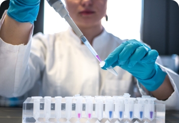
After all the eggs are gathered, they are transferred to a laboratory where a team of embryologists will work on the next stage of the process. Plastic culture dishes have been prepared hours earlier and the eggs are placed into tiny drops of medium, covered in mineral oil to avoid media evaporation. Meanwhile, the man is solicited to supply a semen sample and encouraged to collect this by masturbation in specially designed rooms containing pornographic magazines and/or videos. If he objects, a sample can be brought to the lab before the egg retrieval, collected with a non-toxic condom during intercourse. For obvious reasons, this introduces more variables into the procedure and since personal morals are sometimes subjugated to science, it is often discouraged. While the eggs wait in an incubator and go through the final stages of maturation, the man’s sperm undergoes a series of washes to remove the seminal plasma, non-motile sperm, and other cellular debris using a centrifuge. Key nutrients and proper pH of the culture medium maximize the sperm’s capacity for fertilization within a couple hours of incubation. The embryologist then prepares for insemination of the eggs, performed by one of two possible methods.
- Insemination of the eggs
Prior semen testing and/or IVF attempts will indicate what method of fertilization will be employed. If there is any doubt that the man’s sperm can fertilize the egg, even when placed in immediate proximity, a method called intracytoplasmic sperm injection (ICSI) will be used. Introduced in the early 1990s, ICSI first requires that all the follicular cells attached to the egg are mechanically removed with the help of enzymes that remove the intracellular connections between the cells surrounding each ovum. Then a single sperm is injected deep into the cytoplasm of the egg using micromanipulation equipment and microscopic glass tools. It is a remarkably efficacious method, but invasive, bypassing all the evolutionary mechanisms designed to select the most capable sperm. There are several hurdles for spermatozoa to overcome in the normal fertilization process that IVF with ICSI removes. If the ICSI method is not necessary, several thousand motile sperm, with the precise number of sperm based on the specimen’s observed characteristics, will be placed into the drops of medium containing one or more eggs. The precise number of sperm used represents another area where the art of the practice is honed to maximize the outcome of normal fertilization while minimizing the chance of multiple sperm penetrating the egg, called polyspermy. All the gametes are then incubated overnight.
- Checking fertilization and growing the embryos in culture
An amazing transformation then takes place from the egg and sperm having their own distinct identities expressed in one set of chromosomes (“haploid”) to their coming together to form a completely unique genetic organism, an event happening within a few hours. At the precise moment that the sperm penetrates the outer shell called the zona pellucida and binds to the plasma membrane of the egg, a choreography of events is activated.[5] For the ICSI’d egg, this cascade starts as soon as the membrane of the egg is penetrated by the glass microtool. After this, the new human organism is called a zygote which contains a complete (called “diploid,” two pairs of chromosomes, one male and one female) and thus a unique set of human genes with all the cellular instructions required to grow and develop through all stages of human existence until death. Even more than a newborn, the pre-implantation embryo is completely dependent upon a nurturing environment to survive, simulated to be a maternal substitute. This is achieved by a proprietary culture medium, a petri dish, and an incubator which is gas-, humidity-, and temperature-controlled.
The embryologist performs the evaluation of fertilization precisely 17-20 hours post-insemination.[6] The technician is looking for two individual pronuclei that encapsulate the chromosomes, one from the sperm and one from the egg, before they combine. The fusion of these pronuclei will represent a new zygote. Outside of this time window, the pronuclei will disappear, making it impossible to confirm normal fertilization. The embryos are then placed into fresh growth media and, over the next several days, they will be taken out of the incubator occasionally to observe their development under the microscope.
The knowledge of how to support embryo growth for five days after fertilization was another incredible feat of science applied industry-wide in the early 2000s.[7] The longer culture period reveals, by the survival of the fittest, which embryos have quicker, cleaner and more even cell division. Their speed of cell division along with how cleanly and evenly they divide gives clues as to whether they are likely to successfully implant and survive through pregnancy. Newer technology is being developed which allows time-lapse videography and artificial intelligence to decide which embryos earn the top bid for transfer into the patient.[8] By day five post-fertilization, the embryos should have formed into a blastocyst – an embryo after the first cellular differentiation into two distinct tissues. One is the primitive extra-embryonic membrane involved in implantation, and the other is mostly the fetus along with additional extra-embryonic tissue. At this stage, other micro-manipulation procedures may be performed on the embryo. Micromanipulation uses a high magnification microscope fitted with finely controlled tools to conduct procedures on the embryo such as biopsy for genetic evaluation (called pre-implantation genetic diagnosis) or assisted hatching (a method of thinning the zona pellucida to facilitate easier implantation).
On day five, one or more of the blastocyst embryos are selected for transfer to the uterus of the woman and the remaining embryos may either be frozen for long-term storage (cryopreservation), discarded in medical waste, or used for research which will destroy the embryos, based on the parents’ consent.
Analyzing statistics from the Society of Assisted Reproductive Technology (SART) in 2019 reveals the reason ART success is dependent upon creating excess embryos: given the odds involved, it enables embryo selection and multiple embryo transfer opportunities to increase the probability of pregnancy. Below is a table created from the SART statistics for one attempted IVF cycle and one embryo transfer from patients using their own eggs (Table 1).[9]
Table 1. Statistics for ART pregnancy and pregnancy loss in 2019 (adapted from SART)
| Age of woman | < 35 | 35 – 37 | 38 – 40 | 41 – 42 | > 42 |
| Chemical pregnancy* rate per IVF cycle started | 55.1% | 44.8% | 32.9% | 19.1% | 8.5% |
| Clinical pregnancy* rate per IVF cycle started | 47.5% | 38.3% | 27.5% | 15.5% | 6.3% |
| Live births per IVF cycle started | 40.7% | 31.7% | 22.1% | 11.7% | 3.9% |
| Loss per clinical pregnancy† | 12.7% | 15.3% | 18.0% | 22.2% | 35.0% |
| Loss per chemical pregnancy† | 26.1% | 29.2% | 32.8% | 38.7% | 54.1% |
| Loss per embryo transferred‡ | 55.0% | 59.0% | 69.0% | 80.0% | 93.0% |
*See definitions and discussion of “chemical” and “clinical” pregnancy below.
†These statistics are calculated by subtracting live births from clinical/chemical pregnancies, normalized to those pregnancies. Loss per clinical pregnancy exceeds the SART published miscarriage rate as it encompasses all forms of pregnancy loss after ultrasound confirmation.
‡This statistic is calculated by subtracting live births from the total number of embryos transferred per age group, normalized to that total number transferred.
Focusing on pregnancy loss and embryo mortality changes the perception of IVF success. For example, summarizing the rates for the middle-aged group above, we can say that 32.9% of women 38-40 years old will achieve a positive hCG pregnancy test, 27.5% will achieve a clinical pregnancy, and 22.1% will have a live birth. This means that 32.8% of chemical pregnancies and 18% of clinical pregnancies will result in a loss. Ultimately, 69% of embryos transferred will die in utero. The rates follow a very consistent pattern where the oldest, often unhealthier, patients not only have lower pregnancy rates but also higher losses.
Embryo survival summarized for all age groups and including subsequent frozen embryo transfers reveals a startling statistic: in analyzed SART data from 2019, only an estimated 4-8% of embryos created in the lab were born as infants from transfers conducted within 12 months of retrieval. This low percentage is considered normal despite years of protocol adaptations to increase the likelihood that a given patient will achieve a live birth. An excess number of “spare” embryos are routinely created, leading to their destruction (by discarding in medical waste or by terminal research) or indefinite freezing. This strategy is practically required to ensure even a moderate chance of success and cost-effectiveness, but it is not therapeutic to the embryos created; in fact, it is hostile.
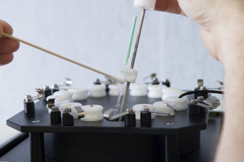
Blastocyst stage embryos are also the stage subjected to freezing for later use, called cryopreservation. Cryopreservation requires that the embryos are first dehydrated and saturated with a cryoprotectant. Examples of cryoprotectants used include the sugar alcohol called glycerol or a diol such as propylene glycol. The fast-freezing method called vitrification is most often used today, which limits intercellular ice crystal damage to the cells. Since methods have evolved, however, embryos in storage today could have been frozen in one of a wide variety of procedures. Therefore, safely thawing them would require knowledge of this evolution of procedures dating back to the 1980s. Thawing requires the stepwise removal of the cryoprotectant in a way that does not explode the cells and kill the embryo. Since the thaw method must be matched to the freeze method (slow freeze, vitrification, etc.), the type of cryoprotectant (glycerol, propylene glycol, etc.), culture medium (including proprietary products and homemade recipes), and the embryo stage (8 cell or blastocyst), these details must be kept in the patient’s lab records. It also requires the technician to be knowledgeable about how to match the protocols and improvise when all the details are not explicit in the records.
Abandoned and discarded embryos continue to plague the industry. By doing simple calculations using data obtained from 2019 CDC, SART, and other sources, the number of human embryos that are cryopreserved or die before being transferred on an annual basis in the U.S. can be estimated. The number of those embryos who die annually, either through simply being discarded or through destructive experimentation, can be estimated at around 446,607; the number who are cryopreserved can be estimated at around 289,491 (see Figure 1). These estimates, however, are conservative, because they only include patients using their own eggs with one embryo transfer and no other categories such as those using donor eggs. They also do not account for embryos that do not survive cryopreservation or are subsequently donated or discarded after cryopreservation.
An ideal solution to the growing number of embryos on ice would be to limit the number of eggs inseminated in new IVF procedures so that no extra embryos are left behind. But this will take outside legislative effort, because there is too much incentive to continue creating excess embryos. As discussed above, success improves by having more embryos to choose from and every frozen embryo is a potential source of revenue for an ART program. The IVF industry would not be unaffected by such regulations, however: it is reasonable to predict that success rates for IVF will decrease if fewer eggs are inseminated until a paradigm shift occurs in the technical approach emphasizing a focus on identifying and selecting the highest quality eggs and sperm, which can be influenced by the health of the parents.
Figure 1.

Data and estimates contributing to this figure are as follows:
* 2019 SART National Summary Report: patients using their own eggs, one embryo transfer
† A total of 1,047,429 oocytes (number of retrievals multiplied by an average of nine oocytes per retrieval [Sunkara et al., 2011 Hum Reprod 26(7)]),[10] multiplied by an estimated 80% fertilization rate (author’s experience)
‡ Initiated cycles with cryopreservation(*), multiplied by an estimated six embryos remaining to freeze per cycle (drawn from assumption of nine oocytes and 80% fertilization rate above, less the mean number of embryos transferred per cycle) and multiplied by 50% embryos judged suitable to freeze (author’s experience)
- Preparation of the woman for embryo transfer and pregnancy monitoring
Transfer is routinely done on day five post-egg retrieval. Recall that most women will have had, just five days earlier, about 10-20 times the normal number of eggs ovulated. The patient will therefore have a very high level of estrogen compared to normal. To offset this and help her uterus be prepared for implantation, she might need supplemental hormones, such as the pregnancy-supporting hormone progesterone.
If everything goes well, one or more of the best appearing embryos (embryos that visually appear healthy) previously selected will be loaded into a catheter and transferred through the cervix into the uterus on day five post-fertilization. Today, over 90% of U.S. live births resulting from ART are singletons. The recommended practice of transferring only one embryo at a time came as a self-correction by the industry to the older norm of transferring 3-5 embryos. The latter resulted in a profoundly increased rate of adverse twinning or multi-embryo pregnancies. These adverse outcomes can affect both the woman and her children due to more than one embryo developing in her uterus. Potential adverse events are also possible in naturally occurring multiple pregnancies, but IVF seems to make this worse (as will be discussed below). To avoid these adverse outcomes, “selective reduction” —i.e., abortion—has been used, where one or more of the implanted embryos are injected with potassium chloride which kills them instantly, leaving one or two of their siblings to be born. Unfortunately, no official statistics are kept on the number of selective reductions conducted today.
Ten to 14 days following the transfer of embryos, blood is collected and measured for human chorionic gonadotropin (hCG). This hormone is produced by the developing placenta and supports a structure on the ovary called the corpus luteum. The corpus luteum develops from a follicle that just released an egg and produces both progesterone and estradiol, which is important during early pregnancy to maintain a healthy uterine lining. Adequate levels of these hormones are essential for the survival of a pregnancy. A positive hCG result via laboratory assay will provide confirmation that a pregnancy has occurred and is often termed a “chemical pregnancy.” With subsequent increases in this hormone every two days, the pregnancy will progress to the stage when it may be detected via ultrasound. At 3-5 weeks post-transfer, if a gestational sac is seen on ultrasound, a “clinical pregnancy” has been achieved (i.e., a pregnancy that has been confirmed both chemically and through ultrasound). Usually, a follow-up ultrasound to document a fetal heartbeat will occur at around 6-8 weeks before care is handed off to an obstetrician. If pregnancy loss occurs, most natural and IVF pregnancies are lost by this milestone, although this can and does happen later in pregnancy.
Other unintended consequences can affect both the woman and the baby. For the women who conceive through ART, there is a higher incidence of perinatal complications, as demonstrated in multi-publication reviews.[11] When making comparisons to naturally conceived pregnancies, it is important that data focus only on singleton pregnancies from IVF to eliminate the known risks associated with multiples.
In a 2017 prospective cohort study, Qin et al.[12] compared pregnancies resulting in spontaneous births from fertile couples, spontaneous births from subfertile couples (couples diagnosed as infertile but who conceived with minimal intervention, not ART), and IVF singleton births. There was a higher incidence of gestational diabetes, hypertension, placenta previa, premature membrane rupture, anemia, pre-term birth, low birth weight, perinatal mortality, and congenital malformations in IVF pregnancies resulting in singleton live birth compared to the pregnancies of fertile couples resulting in live birth. There was an increase in some of the adverse outcomes in the subfertile group compared to the fertile group, but the IVF group also had an even higher incidence of adverse events compared to the subfertile group. The conclusion was that infertility itself leads to some adverse outcomes, but using IVF appears to make these risks worse. This will be explained in more detail below.
Parents who conceive using ART can expect their children to have a 30-40% higher risk of congenital malformations.[13] These anomalies can affect the nervous, craniofacial, heart, genitourinary, gastrointestinal, and musculoskeletal systems.[14] Most birth size abnormalities from ART involve being born small for gestational age, but a subset of infants are born abnormally large, mostly from embryos previously frozen.[15] This introduces the well-known but poorly understood reality of epigenetic changes to the embryo. Epigenetic changes are alterations in how the genes of the new human are expressed.
Lab technique is still a long way from replacing a healthy mother’s body. During the first five days of embryonic life, the embryo normally exists inside the fallopian tube of the mother. This is a dynamic environment where chemical signals, called growth factors, are released by both the epithelium of the fallopian tube and the embryo. This facilitates interactive communication which promotes timely activation of genes that control embryo cell growth. Evolution of the current embryo culture medium started with the removal of nutrients normally used to culture somatic (adult body) cells because they were found to be harmful to embryo growth. Nutrients were then added back when they were subsequently found to promote embryo development.[16]
The media products used today are characterized as being simple and minimal and the ingredients can change slightly based on the stage of embryo development.[17] Despite the huge improvements made to the IVF culture environment over the years, however, there is much more we still don’t understand. In-vitro conditions are much more static than the natural environment, and they are missing many of the factors present in the mother’s fallopian tubes.
In addition to the potential sub-optimal culture conditions compared to the in-vivo environment and the more invasive manipulation of the embryos, harm appears to possibly come from the abnormal maternal environment caused by ovarian stimulation of multiple follicles.[18] Finally, the parent’s underlying health issues are also a factor adding more reasons not to bypass subtle health issues before conception.[19] All of these factors can manifest as cardiometabolic health issues affecting the IVF conceived person later in life.[20]
A Brief History of How IVF Came to Be
Few understand the amount of work that led to the birth of the first baby conceived by IVF, Louise Brown, in 1978. Louise is now a household name, and her birth was the result of research done by the embryologist Robert Edwards, the obstetrician-gynecologist Patrick Steptoe, and nurse/laboratory assistant Jean Purdy. Jean died in 1985 and Dr. Steptoe died in 1988, while Dr. Edwards lived until 2013 and was credited for the achievement by being awarded the Nobel Prize in 2010.
Prior to this success, thousands of research discoveries were made in reproductive science. First, we learned about reproductive anatomy, followed by the discovery of spermatozoa in 1677[21] and the “yellow fleck” discovered to be the first evidence of an oocyte (egg) in 1827.[22] The important discoveries of the various hormones followed later. As early as 1891, Walter Heape produced live rabbits by depositing embryos from one rabbit into another.[23] This kicked off a steady race by scientists to be the first to achieve live births from embryo transfer in other species with or without first using IVF, and earn commensurate academic accolades. The first reports of fertilization in vitro, in this case on rabbits, were published in 1934 by Gregory Pincus and E.V. Enzmann, making big news.[24] In 1944, John Rock reported in vitro fertilization of human eggs.[25] But even though these reports made significant contributions to the field, both “first” titles were stripped from them when it was discovered that sperm must go through a biochemical transformation called capacitation before they can fertilize.[26] Neither Pincus’ nor Rock’s research accounted for this and since eggs can spontaneously begin to divide, a process called parthenogenesis, it cast doubt on those achievements. One of the sperm capacitation discovery scientists, M.C. Chang, immigrated to the U.S. to join Pincus at the Worcester Foundation of Experimental Biology in Massachusetts and in 1959 he was recognized for achieving the first successful mammalian IVF using rabbits.[27]
In 1963, the young scientist Robert Edwards moved to Cambridge to start his own research group funded by the Ford Foundation, and he remained ambitious to pursue human IVF.[28] There he teamed up with Barry Bavister who had learned the critical secret to overcoming one of the hurdles to human IVF, capacitation of spermatozoa.[29] Finally, in 1969, 25 years after Rock’s premature announcement, Robert Edwards achieved the title of first scientist to produce human embryos in vitro. He co-authored the landmark paper in the journal Nature with collaborators Bavister and Steptoe.[30] In Edwards’ report, he describes 56 human eggs being inseminated, but the source of the eggs and spermatozoa are not revealed. In his book, A Matter of Life, however, we learn the details. He explains that the ovarian tissue resulting in a batch of eggs described in the groundbreaking paper came from a woman having her ovary removed.[31] From this tissue they were able to collect 12 eggs which needed to be matured before insemination. After maturing the eggs, he explains that “[t]here was no opportunity in Cambridge of obtaining sperms … so again we collected our own spermatozoa … added them to nine of the 12 eggs, leaving three as controls, and plac[ed] them all in Barry’s culture medium.” Earlier in the book, Edwards describes how his wife responded when he told her his intentions of using his own sperm in the experiments. Her response was “Best of luck.” The women who donated their ovarian tissue knew it was “for research purposes,” but did not know that researchers were attempting to fertilize their eggs with their own sperm and then have the subsequent embryos lethally fixed in acetic acid and stained for later evaluation. One may wonder what they might have thought about this had they known.
These earlier experiments were amazing scientific achievements which led the way for the application of human IVF and to the first human live births,[32] although the ethics of fathering the embryos for destructive embryo research was questionable (to say the least). Much was still needed to be worked out, not least of which was the method of acquiring the eggs from the women. This was the expertise of Patrick Steptoe, who had perfected the use of the laparoscope for aspirating follicles.[33] The technical and scientific challenges required 10 more years of experimentation. It included 282 women; 495 attempted egg collections; 1,361 eggs collected; and 221 embryos described, resulting in 112 embryo transfer attempts. These attempts resulted in 11 pre-clinical early pregnancy losses, one ectopic loss, two miscarriages, and two live births.[34], [35] Clearly, IVF had a long way to go to be clinically relevant, but the spark was created.
The State of IVF Today
IVF procedures today have substantially improved since the first procedures conducted by Steptoe and Edwards. The most recent CDC statistics indicate that 238,126 patients underwent 413,776 IVF cycles at 453 IVF clinics in the U.S. in 2021.[36] Of these cycles, 41% or 167,689 were performed to freeze and store eggs or embryos for later use. From the remaining 246,087 cycles, there were 91,906 live births (some were multiples). This equates to a 37.3% overall chance of live birth. The CDC also reports the success rate on the 144,773 cycles that came from patients using their own eggs and the 24,975 cycles that used donor eggs or embryos. For patients using their own eggs, success rates per attempted retrieval ranged from a high of 50.8% for women <35 years of age to a low of 7.9% for women >40 years of age. Restricting this subset to new patients and including multiple attempted retrievals yields a range from 61.6% to 13.3% for each age group, respectively. Unlike patients using their own eggs, in which cycles may not result in embryos for transfer, patients using donated embryos are guaranteed to have an embryo transfer; therefore, statistics are presented per transfer rather than per attempted retrieval. Using eggs and subsequent embryos that have not been frozen, 53.5% of such transfers result in a live birth (Table 2).
Table 2. Live birth rates in 2021 per retrieval attempt compared to multiple retrieval attempts for IVF (patients’ own eggs) by age and donor egg.
| Egg Source | Live Birth Rate | |||
| Donor egg per transfer | 53.5% | |||
| Age | <35 | 35-37 | 38-40 | >40 |
| Own eggs per attempted retrieval | 50.8% | 36.4% | 23.4% | 7.9% |
| Own eggs from new patients including multiple attempted retrievals | 61.6% | 49.6% | 35.2% | 13.3% |
While success continues to depend on the age of the woman who provides the eggs, as well as other factors, these outcomes are still much better than the < 1% success rate first achieved in 1978. This demonstrates how a strong international effort and financial support can remedy very poor initial outcomes; if the same effort is put into alternatives (discussed below), it just might find more holistic solutions that do not involve the creation of excess human embryos.
IVF and Infertility
At the time of the work of Edwards and Steptoe, achieving pregnancy despite fallopian tube blockage was one possible use of the new technology, but since many reasons for infertility were yet to be described, there was a need to understand just how the new discovery could be used. In Edwards’ Nature paper, discussion of the clinical use of human embryos appeared at the end, where it was claimed that “[f]ertilized human eggs could be useful in treating some forms of infertility.”[37] Subsequently, due to the excitement and interest in this new technology, the numerous pathologies causing infertility needed to conform to the treatment instead of designing technology that targeted each form of pathology.
Today, the CDC publishes the reasons patients pursue assisted reproductive technology. In 2021, the most current statistics, these reasons were diverse and ranged from male infertility to ovarian dysfunction, to uterine tubal occlusion, to recurrent pregnancy loss.[38] This raises the question, if there are multiple reasons for infertility, why is there just one prevailing “solution” for all of them? Why is a diagnosis even needed when the same treatment is going to be used regardless of the pathology? The entire list and frequency of each of these reasons are reproduced below.
Table 3. Reasons for using ART in 2021, related to infertility[39]
|
|
|
|
|
|
|
|
|
The fact that a diagnosis is not really needed is evidenced by the large number of patients labeled as having “other” or “unexplained” factors yet who are still offered ART. Essentially, patients are only diagnosed with what is found, and finding something is a result of how hard someone is looking. Much of the time, the use of ART thus fits the aphorism, “When you have a hammer, everything looks like a nail.” A contrasting observation can be made by looking at the literature published by family physicians who treat patients with infertility without the use of ART. In one such study published in 2021, one observes a change in the diagnosis from “unexplained” to higher incidences of male factor, ovulatory dysfunction, endometriosis, anatomical factors, hypothyroidism, and other endocrine or metabolic diseases. Also included are causes not typically diagnosed in infertility clinics preceding the use of ART, such as limited cervical mucus and premenstrual syndrome.[40]
The CDC also lists reasons for using ART not necessarily related to infertility, reproduced in the following table[41]:
Table 4. Reasons for using ART in 2021, not related to infertility
|
|
|
|
ART has generated these other uses because they clearly did not exist as problems in need of a solution in 1978. They can instead be attributed to changing cultural norms enabled by technology. Today, parents can screen for genetic traits ranging from serious disease to the preferred sex and eye color of their baby. Surrogacy is an added option, even if the woman is completely capable but doesn’t want to carry the pregnancy or experience the pain of delivery/C-section. The American Society for Reproductive Medicine (ASRM) has crafted a definition of “infertility” that would encapsulate the use of ART “regardless of relationship status or sexual orientation.” [42]
Egg and embryo banking, which enable delaying childbearing for career or financial management reasons in no way essentially connected to infertility, are now the largest reasons for using ART. Some may believe this is a positive development, but others might object to the provision of government or insurance coverage for these procedures if used for elective reasons, which need to be teased out of any funding mandate. Corporations are free to provide these benefits, although this could be recognized as a subtle form of coercion to delay family building for job performance purposes.
The roller coaster of emotion
Infertility can be a very painful experience, especially when the chances of ameliorating it are largely beyond the couple’s control—a reality clearly demonstrated by the expressions found on infertility blog sites. A woman on one such site laments, “It was emotionally, mentally and physically draining but I kept going because I wanted a baby. I wanted to be a mom. I wanted to be pregnant and that’s all that mattered to me. Infertility had consumed my every thought and not much else mattered.”[43] These emotions can sometimes be exacerbated by the IVF treatment regimen where each step of the procedure can result in either a high or a low emotional state depending on the outcome. The outcome is contingent on factors like the number of eggs retrieved, fertilization rate, embryos remaining after culture, etc. The most devastating experience is a positive pregnancy test, followed by a pregnancy loss. It reminds us that success encompasses much more than the result of a baby, but also the long-term health and wellbeing of the couple and their relationship. Methods and support ideally need to be compassionate and therapeutic, regardless of the result, and they should never exploit the desperate desire these couples have.[44]
Regulation of IVF in the United States
The advancement of IVF has largely proceeded without significant legislative oversight, except for the technical aspects of the laboratory operation. In the early 1990s, the American Society for Reproductive Medicine (ASRM) and interested groups such as the American Association of Bioanalysts (AAB) took the approach that well-defined self-regulation would negate the need for more formal legislation. Efforts focused on the laboratory and utilized existing legislation designed to regulate other types of clinical laboratory services.[45] Laboratories that provide IVF and other ART procedures typically also provide andrology (such as semen testing) and hormone assay services.
In the early 1990s, if the laboratory was in a hospital, the hormone and andrology procedures would have already been regulated by rules such as those provided in the Clinical Laboratory Improvement Amendments of 1988 (CLIA-88) and by the Occupational Safety and Health Administration (OSHA).[46] Laboratories were also subject to accreditation and inspection by the Joint Commission on Accreditation of Healthcare Organizations (JCAHO, now “The Joint Commission”) and the College of American Pathologists (CAP). In 1992, the Fertility Clinic Success Rate and Certification Act (FCSRCA) and CLIA-88 final rules were enacted, bringing embryology laboratories and physician office laboratories under greater scrutiny. FCSRCA required that all ART programs report success rates to the Department of Health and Human Services (HHS) for public distribution and charged HHS with developing a model program for laboratory accreditation. The CLIA rules also required all laboratories that examine human specimens for the diagnosis, prevention, or treatment of any disease or impairment of the health of human beings to be accredited.
In response, ASRM, partnering with CAP, formed the Reproductive Laboratory Accreditation Program (RLAP). The accreditation rules set standards for the training and experience of personnel, quality control, proficiency, safety, and record keeping. The AAB then created a certification process for embryology and andrology lab personnel.[47] The teeth in these programs include the specimen proficiency testing program and the on-site peer inspection. These programs exert a significant regulatory burden on laboratories regarding their technical and administrative competency. But the efforts by some in the field, mostly by the embryologists and andrologists,[48] to bring ART labs completely under CLIA was never implemented by HHS, leading to less than enforceable regulations for most of the embryology procedures.[49] Similar to the research practices of Edwards and Bavister mentioned above, this leads to precarious conditions for human embryos created in the laboratory.
Other aspects of ART are guided by ethical opinions and practice documents published by ASRM. These publications steer the healthcare practices of thousands of physician members. ASRM is also very active with local legal and legislative activities, pushing not only for IVF access but also for abortion and other issues.[50]
Today’s laboratories are being bought up by large equity firms, increasingly making babies a consumer product that can be chosen like one chooses a home, a car, or a vacation. Corporate culture and funding enable women to put fertility on ice or parents to use genetic screening to ensure the sex and traits of their dream child.[51] Could this be the reason some think the industry is like the “wild west”?[52]
Alternatives and Future Needs: Is IVF the best we can do?
The most significant unintentional outcome of ART may be its suppression of research into and development of alternative methods to diagnose and treat infertility. One example of this is the Estes procedure, used to overcome blocked fallopian tubes until the 1970s when it was strongly criticized by an article published in the IVF journal Fertility and Sterility.[53] We will never know how sophisticated this procedure could have become with all the advancements in non-invasive and robotic surgery now available because it was abandoned for IVF.
Another example, and probably the most vivid demonstration of how IVF suppressed other research, was what happened to male infertility treatment once intracytoplasmic sperm injection (ICSI) swept into the industry.[54] The industry very practically concluded that even knowing why the man is infertile is futile because we don’t know how to treat it. Instead, the woman bears the burden of treatment for the man’s deficiency. Some are questioning the aggressive use of this invasive technique due to the potential consequences including an alteration in the proportion of females born.[55] Imagine if the same effort put forward by Edwards and Steptoe to succeed with IVF were applied to alternatives for resolving male infertility today.
These scenarios have spawned a new and emerging field of medicine called restorative reproductive medicine (RRM).[56] The premise of RRM is that sustained treatments for obvious or even subtle underlying health issues can reverse the cause of infertility. Since there is rarely just one reason for infertility, treatments must be multi-factorial which can take time and become complicated. The fellowship-trained reproductive endocrinologist (RE) is considered the top gun of infertility medicine. Reproductive endocrinology programs train doctors to perform ovarian stimulations, egg retrieval, and embryo transfer, but not typically restorative methods. Patients may assume that all prerequisite testing and treatment prior to application of IVF has been exhausted, but is that true? This brings up the more subtle but important question of motivation. With a multimillion-dollar budget consisting of state-of-the-art laboratory equipment and highly paid laboratory and clinical experts, what incentive is there to try to avoid using the technology? Is this why all roads lead to IVF? Mandated coverage of ART as the primary method of treating infertility may make over-aggressive use even worse.
Although RRM is an emerging field of medicine, there are a few individuals who have been working on RRM for over 30 years and the results are encouraging.[57], [58] The hallmark of the restorative approach is the use of science-based methods of charting the menstrual cycle so that precise timing of blood testing, ultrasound, and hormone treatments can be applied.[59] Examples demonstrate that patients with complicated infertility issues, such as those for whom IVF had previously failed, can be helped.[60], [61] Comparing U.K. IVF data from 2019 to success from an experienced RRM clinic in Dublin, Ireland demonstrates a higher live birth rate compared to a single IVF procedure.[62] Restorative protocols also cost a fraction of a single IVF procedure, and even if couples don’t conceive, they at least have the benefit of improvements to their health.
A shift in the funding for research would enable the tedious but important work of identifying the multiple underlying causes of infertility and cataloging how these abnormalities can be impacted. A reevaluation of the NIH funding budget for reproductive research may be prudent since IVF does not treat the underlying issues and presents a host of unresolvable ethics and regulatory problems; restorative reproductive medicine, though underexplored, provides promise and is deserving of far more support than it currently receives.
Dr. Craig Turczynski completed a B.S in Animal Science at Iowa State University, a Ph.D. in Physiology of Reproduction at Texas A&M University, and a post-doctoral fellowship in reproductive endocrinology at the Women’s Research Institute in Wichita, KS. He became Assistant Professor of ObGyn at Louisiana State University Medical School-Shreveport where he developed and directed their clinical hormone, andrology, and IVF laboratories. After seven years he left the IVF field due to conflicts with the aggressive, life-denying use of the technology and went to work in the medical device industry. Since 2018, he has been rekindling his clinical science career in the field of restorative reproductive medicine by working with leaders in this developing field.
[1] G. Pincus and E. V. Enzmann, “Can Mammalian Eggs Undergo Normal Development in Vitro?,” Proceedings of the National Academy of Sciences of the United States of America 20, no. 2 (February 1934): 121–22, https://doi.org/10.1073/pnas.20.2.121.
[2] Centers for Disease Control and Prevention. “2021 Assisted Reproductive Technology Fertility Clinic and National Summary Report.” US Dept of Health and Human Services; 2023. Available at: https://www.cdc.gov/art/reports/2021/pdf/Report-ART-Fertility-Clinic-National-Summary-H.pdf.
[3] Deekshya Shrestha, Xiaolin La, and Huai L. Feng, “Comparison of Different Stimulation Protocols Used in in Vitro Fertilization: A Review,” Annals of Translational Medicine 3, no. 10 (June 2015): 137, https://doi.org/10.3978/j.issn.2305-5839.2015.04.09.
[4] Practice Committee of the American Society for Reproductive Medicine, “Prevention and treatment of moderate and severe ovarian hyperstimulation syndrome: a guideline.” Fertil Steril. 2016 Dec;106(7):1634-1647. doi: 10.1016/j.fertnstert.2016.08.048.
[5] Nicoletta Tarozzi, Marco Nadalini, Giovanni Coticchio, et al., “The paternal toolbox for embryo development and health,” Molecular Human Reproduction, Volume 27, Issue 7, July 2021, gaab042, https://doi.org/10.1093/molehr/gaab042.
[6] Kobayashi T, Ishikawa H, Ishii K, et al., “Time-lapse monitoring of fertilized human oocytes focused on the incidence of 0PN embryos in conventional in vitro fertilization cycles.” Sci Rep. 2021 Sep 22;11(1):18862. doi: 10.1038/s41598-021-98312-1.
[7] Gardner DK, Vella P, Lane M, et al., “Culture and transfer of human blastocysts increases implantation rates and reduces the need for multiple embryo transfers.” Fertil Steril. 1998 Jan;69(1):84-8. doi: 10.1016/s0015-0282(97)00438-x.
[8] M Salih et al., “Embryo Selection through Artificial Intelligence versus Embryologists: A Systematic Review,” Human Reproduction Open 2023, no. 3 (January 1, 2023): hoad031, https://doi.org/10.1093/hropen/hoad031.
[9] “SART National Summary Report: All Member Clinics,” SART CORS, 2019, https://www.sartcorsonline.com/rptCSR_PublicMultYear.aspx?reportingYear=2019#patient-first-attempt.
[10] Sesh Kamal Sunkara et al., “Association between the Number of Eggs and Live Birth in IVF Treatment: An Analysis of 400 135 Treatment Cycles,” Human Reproduction (Oxford, England) 26, no. 7 (July 2011): 1768–74, https://doi.org/10.1093/humrep/der106.
[11] See for ex.: Sullivan-Pyke CS, Senapati S, Mainigi MA, et al., “In Vitro fertilization and Adverse Obstetric and Perinatal Outcomes.” Semin Perinatol. 2017 Aug 14;41(6):345-353. doi: 10.1053/j.semperi.2017.07.001; Wennerholm UB, Bergh C. “Perinatal outcome in children born after assisted reproductive technologies.” Ups J Med Sci. 2020 May;125(2):158-166. doi: 10.1080/03009734.2020.1726534.
[12] Qin J, Sheng X, Wu D, et al., “Adverse Obstetric Outcomes Associated With In Vitro Fertilization in Singleton Pregnancies: A Prospective Cohort Study.” Reprod Sci. 2017 Dec.;24(4):595-608. doi: 10.1177/1933719116667229.
[13] Chen M, Heilbronn LK, “The health outcomes of human offspring conceived by assisted reproductive technologies (ART).” J Dev Orig Health Dis. 2017 Aug;8(4):388-402. doi: 10.1017/S2040174417000228.
[14] Wennerholm UB, Bergh C. “Perinatal outcome in children born after assisted reproductive technologies.”
[15] Marino JL, Moore VM, Willson KJ, et al., “Perinatal outcomes by mode of assisted conception and sub-fertility in an Australian data linkage cohort.” PLoS One. 2014 Jan 8;9(1):e80398. doi: 10.1371/journal.pone.0080398.
[16] Gardner DK, Lane M. “Culture of viable human blastocysts in defined sequential serum-free media.” Hum Reprod. 1998 Jun;13 Suppl 3:148-59; discussion 160. doi: 10.1093/humrep/13.suppl_3.148.
[17] Simopoulou M, Sfakianoudis K, Rapani A, et al. “Considerations Regarding Embryo Culture Conditions: From Media to Epigenetics.” In Vivo. 2018 May-Jun;32(3):451-460. doi: 10.21873/invivo.11261.
[18] Sullivan-Pyke CS, Senapati S, Mainigi MA, et al., “In Vitro fertilization and Adverse Obstetric and Perinatal Outcomes.” Semin Perinatol. 2017 Aug 14;41(6):345-353. doi: 10.1053/j.semperi.2017.07.001.
[19] Marino JL, et al., “Perinatal outcomes by mode of assisted conception and sub-fertility in an Australian data linkage cohort.”
[20] Pinborg A, Wennerholm UB, Bergh C. “Long-term outcomes for children conceived by assisted reproductive technology.” Fertil Steril. 2023 Sep;120(3 Pt 1):449-456. doi: 10.1016/j.fertnstert.2023.04.022; Huang JY, Cai S, Huang Z, Tint MT, et al. “Analyses of child cardiometabolic phenotype following assisted reproductive technologies using a pragmatic trial emulation approach.” Nat Commun. 2021 Sep 23;12(1):5613. doi: 10.1038/s41467-021-25899-4.
[21] Sharon T. Mortimer, “Essentials of Sperm Biology,” in Office Andrology, ed. Phillip E. Patton and David E. Battaglia (Totowa, NJ: Humana Press, 2005), 1–9, https://doi.org/10.1007/978-1-59259-876-2_1.
[22] Betteridge KJ. “An historical look at embryo transfer.” J Reprod Fertil. 1981 May;62(1):1-13. doi: 10.1530/jrf.0.0620001.
[23] Ibid.
[24] G. Pincus and E. V. Enzmann, “Can Mammalian Eggs Undergo Normal Development in Vitro?”
[25] Stephen Ruffenach, “Rock-Menkin Experiments,” Arizona State University | Embryo Project Encyclopedia, January 13, 2009, https://embryo.asu.edu/pages/rock-menkin-experiments.
[26] Biggers JD, “IVF and embryo transfer: historical origin and development.” Reprod Biomed Online. 2012 Aug;25(2):118-27. doi: 10.1016/j.rbmo.2012.04.011.
[27] Chang MC, “Fertilization of Rabbit Ova in vitro.” Nature. 1959 Aug 8;184(Suppl 7):466-7. doi: 10.1038/184466a0.
[28] Martin H. Johnson, “Robert Edwards: The Path to IVF,” Reproductive Biomedicine Online 23, no. 2 (August 2011): 245–62, https://doi.org/10.1016/j.rbmo.2011.04.010.
[29] Ibid.
[30] Edwards RG, Bavister BD, Steptoe PC, “Early Stages of Fertilization in vitro of Human Oocytes Matured in vitro.” Nature. 1969 Feb 15;221(5181):632-5. doi: 10.1038/221632a0.
[31] Edwards and Steptoe (1980), A Matter of Life: The Story of a Medical Breakthrough. William Morrow and Company; First American Edition. ISBN-10:0688036988; ISBN-13:978-0688036980.
[32] Steptoe PC, Edwards RG, “Birth after the reimplantation of a human embryo.” Lancet. 1978 Aug 12;2(8085):366. doi: 10.1016/s0140-6736(78)92957-4.
[33] Johnson, “Robert Edwards: The Path to IVF.”
[34] Elder K, Johnson MH, “The Oldham Notebooks: an analysis of the development of IVF 1969-1978. II. The treatment cycles and their outcomes.” Reprod Biomed Soc Online. 2015 Jun 14;1(1):9-18. doi: 10.1016/j.rbms.2015.04.003.
[35] Johnson MH, “IVF: The women who helped make it happen.” Reprod Biomed Soc Online. 2018 Dec 14;8:1-6. doi: 10.1016/j.rbms.2018.11.002.
[36] Centers for Disease Control and Prevention. “2021 Assisted Reproductive Technology Fertility Clinic and National Summary Report.”
[37] Edwards RG, Bavister BD, Steptoe PC, “Early Stages of Fertilization in vitro of Human Oocytes Matured in vitro,” p. 635.
[38] Centers for Disease Control and Prevention. “2021 Assisted Reproductive Technology Fertility Clinic and National Summary Report,” p. 17
[39] Percentages sum to greater than 100% because multiple reasons/diagnoses are possible.
[40] Stanford JB, Carpentier PA, Meier BL, et al., “Restorative reproductive medicine for infertility in two family medicine clinics in New England, an observational study,” BMC Pregnancy Childbirth. 2021 Jul 7;21(1):495. doi: 10.1186/s12884-021-03946-8.
[41] Centers for Disease Control and Prevention. “2021 Assisted Reproductive Technology Fertility Clinic and National Summary Report,” p. 17.
[42] “Definition of Infertility: A Committee Opinion” (Practice Committee of the American Society for Reproductive Medicine, 2023), https://www.fertstert.org/action/showPdf?pii=S0015-0282%2823%2901971-4.
[43] “A Story of Infertility: A Childless Ending,” Infertility Unfiltered, accessed November 15, 2024, https://www.infertilityunfiltered.com/blog/2022/8/15/a-story-of-infertility-childless.
[44] The Lancet, “The fertility industry: profiting from vulnerability,” The Lancet 2024 Jul 20;404(10449):215. doi: 10.1016/S0140-6736(24)01484-3.
[45] Gerrity M., “Legislative efforts affecting the reproductive biology laboratory,” Curr Opin Obstet Gynecol 1993 Oct;5(5):623-9, https://pubmed.ncbi.nlm.nih.gov/8241438/.
[46] Gerrity M., “Legislative efforts affecting the reproductive biology laboratory,” Curr Opin Obstet Gynecol 1993 Oct;5(5):623-9, https://pubmed.ncbi.nlm.nih.gov/8241438/.
[47] Ibid.
[48] American Association of Bioanalysts, “History of the College of Reproductive Biology (CRB),” September 29, 2016; accessed November 26, 2024, https://www.aab.org/images/History%20of%20the%20CRB%20updated%209-29-16.pdf.
[49] Ibid.; See also Mary Harned, “IVF Industry Regulation in the United States: Changes Are Needed to Protect Embryonic Children and their Families,” Charlotte Lozier Institute, On Point Series 101, accessed November 26, 2024, https://lozierinstitute.org/ivf-industry-regulation-in-the-united-states-changes-are-needed-to-protect-embryonic-children-and-their-families/
[50] “2024 Advocacy Activities,” American Society for Reproductive Medicine, accessed November 19, 2024, https://www.asrm.org/advocacy-and-policy/advocacy-activities/2024-advocacy-activities/.
[51] Emi Nietfeld, “The Parents Who Want Daughters–and Daughters Only,” Slate, May 7, 2024, https://slate.com/technology/2024/05/ivf-daughters-toxic-masculinity-sex-selection.html.
[52] Susan Crockin and Francesca Nardi, “Alabama Supreme Court Rules Frozen Embryos Are ‘Unborn Children’ and Admonishes IVF’s ‘Wild West’ Treatment,” American Society for Reproductive Medicine, accessed November 22, 2024, https://www.asrm.org/news-and-events/asrm-news/legally-speaking/frozen-embryo-destruction-and–potential-travel-restrictions-for-surrogacy-arrangements2/.
[53] Beyth Y, Polishuk WZ, “Ovarian implantation into the uterus (Estes operation): clinical and experimental evaluation.” Fertil Steril. 1979 Dec;32(6):657-60. doi: 10.1016/s0015-0282(16)44414-6.
[54] Palermo G, Joris H, Devroey P, Van Steirteghem AC, “Pregnancies after intracytoplasmic injection of single spermatozoon into an oocyte.” Lancet. 1992 Jul 4;340(8810):17-8. doi: 10.1016/0140-6736(92)92425-f.
[55] Edgardo Somigliana, Giussy Barbara, Laila Giorgia Micci, et al. “ICSI for non-male infertility: from ineffectiveness to gender-bias?,” Reproductive BioMedicine Online, 2024, 104706, ISSN 1472-6483, https://doi.org/10.1016/j.rbmo.2024.104706.
[56] “Home,” International Institute for Restorative Reproductive Medicine, accessed November 19, 2024, https://iirrm.org/.
[57] Stanford JB, Parnell TA, Boyle PC, “Outcomes from treatment of infertility with natural procreative technology in an Irish general practice.” J Am Board Fam Med. 2008 Sep-Oct;21(5):375-84. doi: 10.3122/jabfm.2008.05.070239. Erratum in: J Am Board Fam Med. 2008 Nov-Dec;21(6):583.
[58] Tham E, Schliep K, Stanford J, “Natural procreative technology for infertility and recurrent miscarriage: outcomes in a Canadian family practice.” Can Fam Physician. 2012 May;58(5):e267-74, https://pubmed.ncbi.nlm.nih.gov/22734170/.
[59] Duane M, Stanford JB, Porucznik CA, Vigil P, “Fertility Awareness-Based Methods for Women’s Health and Family Planning.” Front Med (Lausanne). 2022 May 24;9:858977. doi: 10.3389/fmed.2022.858977.
[60] Boyle PC, de Groot T, Andralojc KM, Parnell TA, “Healthy Singleton Pregnancies from Restorative Reproductive Medicine (RRM) After Failed IVF.” Front Med (Lausanne). 2018 Jul 31;5:210. doi: 10.3389/fmed.2018.00210.
[61] Boyle PC, Stanford JB, Zecevic I, “Successful pregnancy with restorative reproductive medicine after 16 years of infertility, three recurrent miscarriages, and eight unsuccessful embryo transfers with in vitro fertilization/intracytoplasmic sperm injection: a case report.” J Med Case Rep. 2022 Jun 22;16(1):246. doi: 10.1186/s13256-022-03465-w.
[62] Boyle, PC, Toth, A, ONeill, L, Turczynski, CJ, “Restorative Reproductive Medicine: An Emerging New Treatment Process and a Prerequisite to Assisted Reproductive Technology for Treatment of Infertility.” Preprints 2024, 2024010624. https://doi.org/10.20944/preprints202401.0624.v1.